{"title":"巨大颈内动脉瘤分流治疗失败后重建夹板手术颅内外搭桥一例报告。","authors":"Hung Manh Ngo, Minh Quang Ngo","doi":"10.1055/s-0045-1805019","DOIUrl":null,"url":null,"abstract":"<p><p>Flow diverter (FD) is often the first-line treatment for giant internal carotid artery aneurysms, with a high rate of aneurysm occlusion. However, up to 10% of giant cerebral aneurysms increase in size after FD treatment. Surgery is usually considered if the giant internal carotid artery aneurysm continues to enlarge and cause compression. We report a case of a giant internal carotid artery aneurysm that continued to increase in size after FD treatment and was subsequently treated surgically. We also review the literature on the management of giant cerebral aneurysms that increased in size after FD. A 41-year-old female patient was diagnosed with a right giant internal carotid artery aneurysm and was initially treated with FD. After FD, the patient's vision in the right eye did not improve. Despite medical treatment, her vision continued to deteriorate. The patient presented at our hospital with reduced vision in both eyes. Magnetic resonance imaging and digital subtraction angiography with balloon test occlusion confirmed the presence of a giant thrombosed aneurysm in the right internal carotid artery, compressing the right optic nerve and optic chiasm. The patient underwent external carotid artery-middle cerebral artery bypass surgery using a radial artery graft, aneurysm sac dissection with thrombus removal, and reconstructive clipping of the aneurysm neck. After surgery, the patient's vision in both eyes improved immediately and did not develop any new neurological symptoms. Extracranial-intracranial arterial reconstructive surgery is a viable option for treating giant internal carotid artery aneurysms that have undergone FD treatment but continue to present with progressive mass effects.</p>","PeriodicalId":94300,"journal":{"name":"Asian journal of neurosurgery","volume":"20 2","pages":"391-396"},"PeriodicalIF":0.0000,"publicationDate":"2025-03-20","publicationTypes":"Journal Article","fieldsOfStudy":null,"isOpenAccess":false,"openAccessPdf":"https://www.ncbi.nlm.nih.gov/pmc/articles/PMC12136965/pdf/","citationCount":"0","resultStr":"{\"title\":\"Extracranial-Intracranial Bypass with Reconstruction Clip Surgery Following Failed Flow Diverter Therapy for a Giant Internal Carotid Aneurysm: A Case Report.\",\"authors\":\"Hung Manh Ngo, Minh Quang Ngo\",\"doi\":\"10.1055/s-0045-1805019\",\"DOIUrl\":null,\"url\":null,\"abstract\":\"<p><p>Flow diverter (FD) is often the first-line treatment for giant internal carotid artery aneurysms, with a high rate of aneurysm occlusion. However, up to 10% of giant cerebral aneurysms increase in size after FD treatment. Surgery is usually considered if the giant internal carotid artery aneurysm continues to enlarge and cause compression. We report a case of a giant internal carotid artery aneurysm that continued to increase in size after FD treatment and was subsequently treated surgically. We also review the literature on the management of giant cerebral aneurysms that increased in size after FD. A 41-year-old female patient was diagnosed with a right giant internal carotid artery aneurysm and was initially treated with FD. After FD, the patient's vision in the right eye did not improve. Despite medical treatment, her vision continued to deteriorate. The patient presented at our hospital with reduced vision in both eyes. Magnetic resonance imaging and digital subtraction angiography with balloon test occlusion confirmed the presence of a giant thrombosed aneurysm in the right internal carotid artery, compressing the right optic nerve and optic chiasm. The patient underwent external carotid artery-middle cerebral artery bypass surgery using a radial artery graft, aneurysm sac dissection with thrombus removal, and reconstructive clipping of the aneurysm neck. After surgery, the patient's vision in both eyes improved immediately and did not develop any new neurological symptoms. Extracranial-intracranial arterial reconstructive surgery is a viable option for treating giant internal carotid artery aneurysms that have undergone FD treatment but continue to present with progressive mass effects.</p>\",\"PeriodicalId\":94300,\"journal\":{\"name\":\"Asian journal of neurosurgery\",\"volume\":\"20 2\",\"pages\":\"391-396\"},\"PeriodicalIF\":0.0000,\"publicationDate\":\"2025-03-20\",\"publicationTypes\":\"Journal Article\",\"fieldsOfStudy\":null,\"isOpenAccess\":false,\"openAccessPdf\":\"https://www.ncbi.nlm.nih.gov/pmc/articles/PMC12136965/pdf/\",\"citationCount\":\"0\",\"resultStr\":null,\"platform\":\"Semanticscholar\",\"paperid\":null,\"PeriodicalName\":\"Asian journal of neurosurgery\",\"FirstCategoryId\":\"1085\",\"ListUrlMain\":\"https://doi.org/10.1055/s-0045-1805019\",\"RegionNum\":0,\"RegionCategory\":null,\"ArticlePicture\":[],\"TitleCN\":null,\"AbstractTextCN\":null,\"PMCID\":null,\"EPubDate\":\"2025/6/1 0:00:00\",\"PubModel\":\"eCollection\",\"JCR\":\"\",\"JCRName\":\"\",\"Score\":null,\"Total\":0}","platform":"Semanticscholar","paperid":null,"PeriodicalName":"Asian journal of neurosurgery","FirstCategoryId":"1085","ListUrlMain":"https://doi.org/10.1055/s-0045-1805019","RegionNum":0,"RegionCategory":null,"ArticlePicture":[],"TitleCN":null,"AbstractTextCN":null,"PMCID":null,"EPubDate":"2025/6/1 0:00:00","PubModel":"eCollection","JCR":"","JCRName":"","Score":null,"Total":0}
Extracranial-Intracranial Bypass with Reconstruction Clip Surgery Following Failed Flow Diverter Therapy for a Giant Internal Carotid Aneurysm: A Case Report.
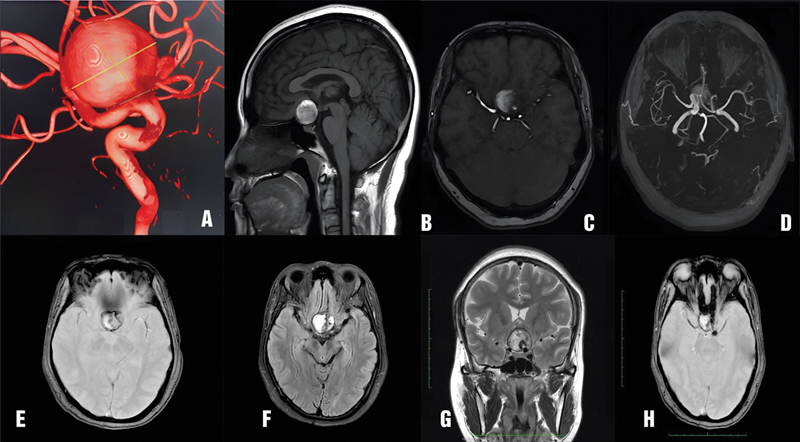
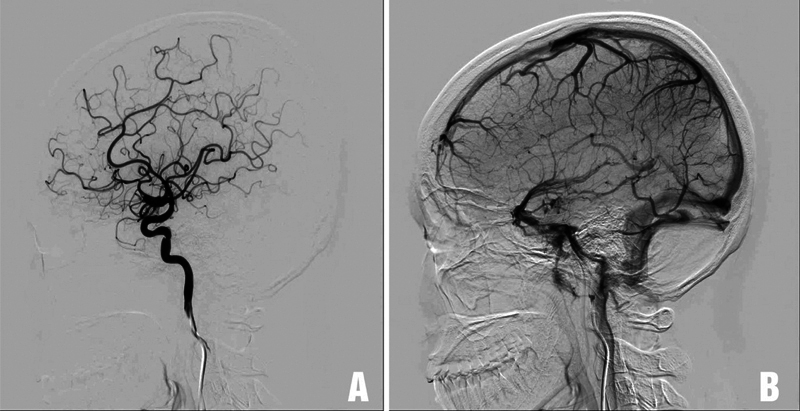
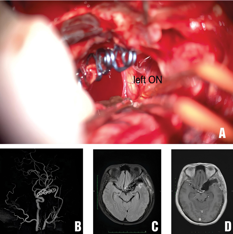
Flow diverter (FD) is often the first-line treatment for giant internal carotid artery aneurysms, with a high rate of aneurysm occlusion. However, up to 10% of giant cerebral aneurysms increase in size after FD treatment. Surgery is usually considered if the giant internal carotid artery aneurysm continues to enlarge and cause compression. We report a case of a giant internal carotid artery aneurysm that continued to increase in size after FD treatment and was subsequently treated surgically. We also review the literature on the management of giant cerebral aneurysms that increased in size after FD. A 41-year-old female patient was diagnosed with a right giant internal carotid artery aneurysm and was initially treated with FD. After FD, the patient's vision in the right eye did not improve. Despite medical treatment, her vision continued to deteriorate. The patient presented at our hospital with reduced vision in both eyes. Magnetic resonance imaging and digital subtraction angiography with balloon test occlusion confirmed the presence of a giant thrombosed aneurysm in the right internal carotid artery, compressing the right optic nerve and optic chiasm. The patient underwent external carotid artery-middle cerebral artery bypass surgery using a radial artery graft, aneurysm sac dissection with thrombus removal, and reconstructive clipping of the aneurysm neck. After surgery, the patient's vision in both eyes improved immediately and did not develop any new neurological symptoms. Extracranial-intracranial arterial reconstructive surgery is a viable option for treating giant internal carotid artery aneurysms that have undergone FD treatment but continue to present with progressive mass effects.

 求助内容:
求助内容: 应助结果提醒方式:


