Xiaoming Sun, Lin Lv, Daming Fan, Yubin Huang, Liangming Zhu, Haibo Liu
{"title":"氟-18-氟脱氧葡萄糖正电子发射断层扫描误诊食管平滑肌瘤合并肾上腺皮质腺瘤为食管癌伴肾上腺转移1例。","authors":"Xiaoming Sun, Lin Lv, Daming Fan, Yubin Huang, Liangming Zhu, Haibo Liu","doi":"10.3892/ol.2025.14984","DOIUrl":null,"url":null,"abstract":"<p><p>Leiomyoma is a benign muscular abnormality that commonly occurs in the middle and distal third of the esophagus, leading to thickening of the esophageal wall and subsequent esophageal luminal narrowing. Notably, esophageal leiomyoma often does not show increased 18F-fluorodeoxyglucose (FDG) uptake on positron emission tomography (PET). The present study described a case of esophageal leiomyoma combined with adrenal adenoma. Results of the PET-computed tomography analysis revealed that FDG metabolism was increased in the lower segment of the esophagus and the left adrenal gland, with maximum standardized uptake values of 6.5 and 4.1, respectively. Therefore, initially, the patient was diagnosed with an esophageal malignant tumor with left adrenal metastasis. Open surgery was performed for complete removal of the lesions, and results of a routine pathological analysis revealed esophageal leiomyoma combined with adrenal cortical adenoma. The present study indicates that to avoid unnecessary surgeries, esophageal leiomyoma and adrenal cortical adenoma should be diagnosed through a comprehensive assessment with endoscopy, endoscopic ultrasound, computed tomography, magnetic resonance imaging and tissue sample pathology, not just PET.</p>","PeriodicalId":19503,"journal":{"name":"Oncology Letters","volume":"29 5","pages":"238"},"PeriodicalIF":2.2000,"publicationDate":"2025-03-20","publicationTypes":"Journal Article","fieldsOfStudy":null,"isOpenAccess":false,"openAccessPdf":"https://www.ncbi.nlm.nih.gov/pmc/articles/PMC12142281/pdf/","citationCount":"0","resultStr":"{\"title\":\"Misdiagnosis of esophageal leiomyoma combined with adrenal cortical adenoma as esophageal cancer with adrenal metastasis by fluorine-18-fluorodeoxyglucose positron emission tomography: A case report.\",\"authors\":\"Xiaoming Sun, Lin Lv, Daming Fan, Yubin Huang, Liangming Zhu, Haibo Liu\",\"doi\":\"10.3892/ol.2025.14984\",\"DOIUrl\":null,\"url\":null,\"abstract\":\"<p><p>Leiomyoma is a benign muscular abnormality that commonly occurs in the middle and distal third of the esophagus, leading to thickening of the esophageal wall and subsequent esophageal luminal narrowing. Notably, esophageal leiomyoma often does not show increased 18F-fluorodeoxyglucose (FDG) uptake on positron emission tomography (PET). The present study described a case of esophageal leiomyoma combined with adrenal adenoma. Results of the PET-computed tomography analysis revealed that FDG metabolism was increased in the lower segment of the esophagus and the left adrenal gland, with maximum standardized uptake values of 6.5 and 4.1, respectively. Therefore, initially, the patient was diagnosed with an esophageal malignant tumor with left adrenal metastasis. Open surgery was performed for complete removal of the lesions, and results of a routine pathological analysis revealed esophageal leiomyoma combined with adrenal cortical adenoma. The present study indicates that to avoid unnecessary surgeries, esophageal leiomyoma and adrenal cortical adenoma should be diagnosed through a comprehensive assessment with endoscopy, endoscopic ultrasound, computed tomography, magnetic resonance imaging and tissue sample pathology, not just PET.</p>\",\"PeriodicalId\":19503,\"journal\":{\"name\":\"Oncology Letters\",\"volume\":\"29 5\",\"pages\":\"238\"},\"PeriodicalIF\":2.2000,\"publicationDate\":\"2025-03-20\",\"publicationTypes\":\"Journal Article\",\"fieldsOfStudy\":null,\"isOpenAccess\":false,\"openAccessPdf\":\"https://www.ncbi.nlm.nih.gov/pmc/articles/PMC12142281/pdf/\",\"citationCount\":\"0\",\"resultStr\":null,\"platform\":\"Semanticscholar\",\"paperid\":null,\"PeriodicalName\":\"Oncology Letters\",\"FirstCategoryId\":\"3\",\"ListUrlMain\":\"https://doi.org/10.3892/ol.2025.14984\",\"RegionNum\":4,\"RegionCategory\":\"医学\",\"ArticlePicture\":[],\"TitleCN\":null,\"AbstractTextCN\":null,\"PMCID\":null,\"EPubDate\":\"2025/5/1 0:00:00\",\"PubModel\":\"eCollection\",\"JCR\":\"Q3\",\"JCRName\":\"ONCOLOGY\",\"Score\":null,\"Total\":0}","platform":"Semanticscholar","paperid":null,"PeriodicalName":"Oncology Letters","FirstCategoryId":"3","ListUrlMain":"https://doi.org/10.3892/ol.2025.14984","RegionNum":4,"RegionCategory":"医学","ArticlePicture":[],"TitleCN":null,"AbstractTextCN":null,"PMCID":null,"EPubDate":"2025/5/1 0:00:00","PubModel":"eCollection","JCR":"Q3","JCRName":"ONCOLOGY","Score":null,"Total":0}
Misdiagnosis of esophageal leiomyoma combined with adrenal cortical adenoma as esophageal cancer with adrenal metastasis by fluorine-18-fluorodeoxyglucose positron emission tomography: A case report.
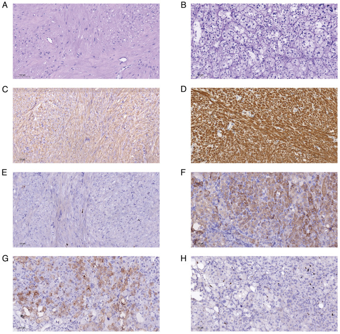
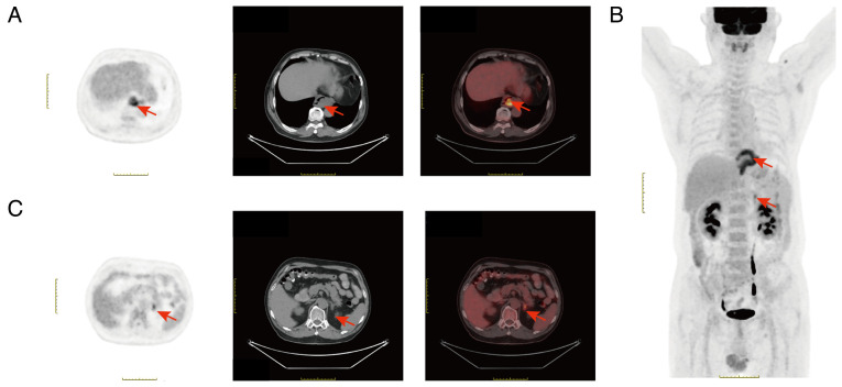
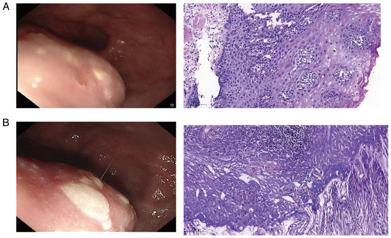
Leiomyoma is a benign muscular abnormality that commonly occurs in the middle and distal third of the esophagus, leading to thickening of the esophageal wall and subsequent esophageal luminal narrowing. Notably, esophageal leiomyoma often does not show increased 18F-fluorodeoxyglucose (FDG) uptake on positron emission tomography (PET). The present study described a case of esophageal leiomyoma combined with adrenal adenoma. Results of the PET-computed tomography analysis revealed that FDG metabolism was increased in the lower segment of the esophagus and the left adrenal gland, with maximum standardized uptake values of 6.5 and 4.1, respectively. Therefore, initially, the patient was diagnosed with an esophageal malignant tumor with left adrenal metastasis. Open surgery was performed for complete removal of the lesions, and results of a routine pathological analysis revealed esophageal leiomyoma combined with adrenal cortical adenoma. The present study indicates that to avoid unnecessary surgeries, esophageal leiomyoma and adrenal cortical adenoma should be diagnosed through a comprehensive assessment with endoscopy, endoscopic ultrasound, computed tomography, magnetic resonance imaging and tissue sample pathology, not just PET.
期刊介绍:
Oncology Letters is a monthly, peer-reviewed journal, available in print and online, that focuses on all aspects of clinical oncology, as well as in vitro and in vivo experimental model systems relevant to the mechanisms of disease.
The principal aim of Oncology Letters is to provide the prompt publication of original studies of high quality that pertain to clinical oncology, chemotherapy, oncogenes, carcinogenesis, metastasis, epidemiology and viral oncology in the form of original research, reviews and case reports.
 求助内容:
求助内容: 应助结果提醒方式:


