Kristof Babarczy, Bence L Radics, Orsolya Horvath, Peter Klivenyi, Levente Szalardy
{"title":"静脉溶栓的致命结果与淀粉样蛋白β相关性血管炎的意外发现-一个病例报告强调了急性局灶性神经功能缺损和最小的影像学表现的相关情况。","authors":"Kristof Babarczy, Bence L Radics, Orsolya Horvath, Peter Klivenyi, Levente Szalardy","doi":"10.1111/neup.70013","DOIUrl":null,"url":null,"abstract":"<p><p>Cerebral amyloid angiopathy (CAA) has been implicated as a risk for developing lobar intracerebral hemorrhage (ICH) after intravenous thrombolysis (IVT) applied for acute ischemic stroke (AIS). However, there is a paucity of cases reported with histopathological CAA diagnosis in this setting, with a single report to imply the role of CAA-related inflammation (CAA-RI). We report clinical, radiological, and neuropathological observations of a 65-year-old woman who presented with acute left-hemispheric symptoms with an initially unrevealing cranial computed tomography (CT) and received IVT for presumed AIS. The course was rapidly complicated by a huge lobar ICH and a fatal outcome. The autopsy revealed severe CAA, unexpectedly with transmural CAA-RI, a.k.a. amyloid-β-related angiitis (ABRA), and histopathological evidence for vascular amyloid-β phagocytosis. Re-evaluation of initial imaging did not reveal signs of asymmetric confluent white matter edema characteristic of CAA-RI, but raised the suspicion of a tiny left central convexity subarachnoid hemorrhage, a substrate of amyloid spells. The genotype of the apolipoprotein E (ApoE) gene (ApoE) was ε3/ε3. Being the second published thrombolysis-associated fatality with ABRA and among the few with definite CAA, the present case confirms CAA/CAA-RI to be a potential hidden risk for IVT-associated ICHs, urging for awareness of CAA-associated pathologies and clinical-radiological hints in an AIS setting. The findings implicate the relevance of vascular Aβ phagocytosis in the pathogenesis, confirm that CAA-RI may present without prominent edema, highlight that CAA/CAA-RI-related focal neurological deficits (including amyloid spells) can be potential AIS mimics within the IVT time window, and urge for rigorous analysis of pre-IVT CT scans for even subtle sulcal hyperdensities suggesting cSAH/amyloid spell in elderly patients, prompting consideration of magnetic resonance imaging.</p>","PeriodicalId":19204,"journal":{"name":"Neuropathology","volume":" ","pages":"e70013"},"PeriodicalIF":1.2000,"publicationDate":"2025-08-01","publicationTypes":"Journal Article","fieldsOfStudy":null,"isOpenAccess":false,"openAccessPdf":"https://www.ncbi.nlm.nih.gov/pmc/articles/PMC12279614/pdf/","citationCount":"0","resultStr":"{\"title\":\"Fatal Outcome of Intravenous Thrombolysis With an Unexpected Finding of Amyloid-β-Related Angiitis-A Case Report Highlighting a Relevant Scenario With Acute Focal Neurological Deficits and Minimal Radiological Presentation.\",\"authors\":\"Kristof Babarczy, Bence L Radics, Orsolya Horvath, Peter Klivenyi, Levente Szalardy\",\"doi\":\"10.1111/neup.70013\",\"DOIUrl\":null,\"url\":null,\"abstract\":\"<p><p>Cerebral amyloid angiopathy (CAA) has been implicated as a risk for developing lobar intracerebral hemorrhage (ICH) after intravenous thrombolysis (IVT) applied for acute ischemic stroke (AIS). However, there is a paucity of cases reported with histopathological CAA diagnosis in this setting, with a single report to imply the role of CAA-related inflammation (CAA-RI). We report clinical, radiological, and neuropathological observations of a 65-year-old woman who presented with acute left-hemispheric symptoms with an initially unrevealing cranial computed tomography (CT) and received IVT for presumed AIS. The course was rapidly complicated by a huge lobar ICH and a fatal outcome. The autopsy revealed severe CAA, unexpectedly with transmural CAA-RI, a.k.a. amyloid-β-related angiitis (ABRA), and histopathological evidence for vascular amyloid-β phagocytosis. Re-evaluation of initial imaging did not reveal signs of asymmetric confluent white matter edema characteristic of CAA-RI, but raised the suspicion of a tiny left central convexity subarachnoid hemorrhage, a substrate of amyloid spells. The genotype of the apolipoprotein E (ApoE) gene (ApoE) was ε3/ε3. Being the second published thrombolysis-associated fatality with ABRA and among the few with definite CAA, the present case confirms CAA/CAA-RI to be a potential hidden risk for IVT-associated ICHs, urging for awareness of CAA-associated pathologies and clinical-radiological hints in an AIS setting. The findings implicate the relevance of vascular Aβ phagocytosis in the pathogenesis, confirm that CAA-RI may present without prominent edema, highlight that CAA/CAA-RI-related focal neurological deficits (including amyloid spells) can be potential AIS mimics within the IVT time window, and urge for rigorous analysis of pre-IVT CT scans for even subtle sulcal hyperdensities suggesting cSAH/amyloid spell in elderly patients, prompting consideration of magnetic resonance imaging.</p>\",\"PeriodicalId\":19204,\"journal\":{\"name\":\"Neuropathology\",\"volume\":\" \",\"pages\":\"e70013\"},\"PeriodicalIF\":1.2000,\"publicationDate\":\"2025-08-01\",\"publicationTypes\":\"Journal Article\",\"fieldsOfStudy\":null,\"isOpenAccess\":false,\"openAccessPdf\":\"https://www.ncbi.nlm.nih.gov/pmc/articles/PMC12279614/pdf/\",\"citationCount\":\"0\",\"resultStr\":null,\"platform\":\"Semanticscholar\",\"paperid\":null,\"PeriodicalName\":\"Neuropathology\",\"FirstCategoryId\":\"3\",\"ListUrlMain\":\"https://doi.org/10.1111/neup.70013\",\"RegionNum\":4,\"RegionCategory\":\"医学\",\"ArticlePicture\":[],\"TitleCN\":null,\"AbstractTextCN\":null,\"PMCID\":null,\"EPubDate\":\"2025/6/5 0:00:00\",\"PubModel\":\"Epub\",\"JCR\":\"Q4\",\"JCRName\":\"CLINICAL NEUROLOGY\",\"Score\":null,\"Total\":0}","platform":"Semanticscholar","paperid":null,"PeriodicalName":"Neuropathology","FirstCategoryId":"3","ListUrlMain":"https://doi.org/10.1111/neup.70013","RegionNum":4,"RegionCategory":"医学","ArticlePicture":[],"TitleCN":null,"AbstractTextCN":null,"PMCID":null,"EPubDate":"2025/6/5 0:00:00","PubModel":"Epub","JCR":"Q4","JCRName":"CLINICAL NEUROLOGY","Score":null,"Total":0}
Fatal Outcome of Intravenous Thrombolysis With an Unexpected Finding of Amyloid-β-Related Angiitis-A Case Report Highlighting a Relevant Scenario With Acute Focal Neurological Deficits and Minimal Radiological Presentation.
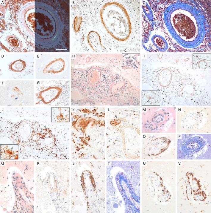
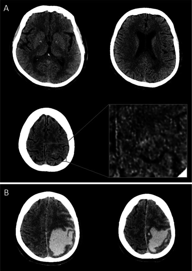
Cerebral amyloid angiopathy (CAA) has been implicated as a risk for developing lobar intracerebral hemorrhage (ICH) after intravenous thrombolysis (IVT) applied for acute ischemic stroke (AIS). However, there is a paucity of cases reported with histopathological CAA diagnosis in this setting, with a single report to imply the role of CAA-related inflammation (CAA-RI). We report clinical, radiological, and neuropathological observations of a 65-year-old woman who presented with acute left-hemispheric symptoms with an initially unrevealing cranial computed tomography (CT) and received IVT for presumed AIS. The course was rapidly complicated by a huge lobar ICH and a fatal outcome. The autopsy revealed severe CAA, unexpectedly with transmural CAA-RI, a.k.a. amyloid-β-related angiitis (ABRA), and histopathological evidence for vascular amyloid-β phagocytosis. Re-evaluation of initial imaging did not reveal signs of asymmetric confluent white matter edema characteristic of CAA-RI, but raised the suspicion of a tiny left central convexity subarachnoid hemorrhage, a substrate of amyloid spells. The genotype of the apolipoprotein E (ApoE) gene (ApoE) was ε3/ε3. Being the second published thrombolysis-associated fatality with ABRA and among the few with definite CAA, the present case confirms CAA/CAA-RI to be a potential hidden risk for IVT-associated ICHs, urging for awareness of CAA-associated pathologies and clinical-radiological hints in an AIS setting. The findings implicate the relevance of vascular Aβ phagocytosis in the pathogenesis, confirm that CAA-RI may present without prominent edema, highlight that CAA/CAA-RI-related focal neurological deficits (including amyloid spells) can be potential AIS mimics within the IVT time window, and urge for rigorous analysis of pre-IVT CT scans for even subtle sulcal hyperdensities suggesting cSAH/amyloid spell in elderly patients, prompting consideration of magnetic resonance imaging.
期刊介绍:
Neuropathology is an international journal sponsored by the Japanese Society of Neuropathology and publishes peer-reviewed original papers dealing with all aspects of human and experimental neuropathology and related fields of research. The Journal aims to promote the international exchange of results and encourages authors from all countries to submit papers in the following categories: Original Articles, Case Reports, Short Communications, Occasional Reviews, Editorials and Letters to the Editor. All articles are peer-reviewed by at least two researchers expert in the field of the submitted paper.
 求助内容:
求助内容: 应助结果提醒方式:


