Elin Bäck, My Jonasson, Elin Lindström, Andreas Tolf, Joachim Burman, Lieuwe Appel, Mark Lubberink
{"title":"图像重建对15o -水正电子发射断层扫描测量脑血流的影响。","authors":"Elin Bäck, My Jonasson, Elin Lindström, Andreas Tolf, Joachim Burman, Lieuwe Appel, Mark Lubberink","doi":"10.1186/s40658-025-00760-5","DOIUrl":null,"url":null,"abstract":"<p><strong>Background: </strong><sup>15</sup>O-water positron emission tomography (PET) is considered the gold standard method for non-invasive measurement of cerebral blood flow (CBF). However, previously published average CBF values in healthy subjects have varied greatly and the cause of these variations remains unclear. This study investigates how image reconstruction methods and spatial resolution affect CBF measurements with <sup>15</sup>O-water PET.</p><p><strong>Methods: </strong>Eight healthy subjects each underwent dynamic <sup>15</sup>O-water PET scans with continuous arterial blood sampling. Images were reconstructed using two different algorithms; ordered subset expectation maximisation and block sequential regularised expectation maximalisation with varying reconstruction parameters. CBF was estimated for the whole brain, grey matter, and central white matter. Reconstruction-specific effective spatial resolution was estimated using phantom measurements and simulations.</p><p><strong>Results: </strong>The mean whole brain CBF was 0.48 mL/cm<sup>3</sup>/min and showed little dependence on the image reconstruction method. Grey matter CBF varied between 0.52 and 0.57 mL/cm<sup>3</sup>/min, and central white matter CBF between 0.20 and 0.28 mL/cm<sup>3</sup>/min. Regional CBF showed great dependence on effective spatial resolution with a negative correlation between grey matter CBF and resolution (r = -0.96) and a positive correlation between central white matter and resolution (r = 0.93).</p><p><strong>Conclusion: </strong>This study concludes that grey matter and central white matter CBF, but not whole brain CBF measured with quantitative <sup>15</sup>O-water PET is reconstruction method dependent, mainly due to varying spatial resolution with consequent partial volume effects. Variations in published CBF values cannot be explained solely by reconstruction methods or spatial resolution.</p>","PeriodicalId":11559,"journal":{"name":"EJNMMI Physics","volume":"12 1","pages":"52"},"PeriodicalIF":3.2000,"publicationDate":"2025-06-06","publicationTypes":"Journal Article","fieldsOfStudy":null,"isOpenAccess":false,"openAccessPdf":"https://www.ncbi.nlm.nih.gov/pmc/articles/PMC12144024/pdf/","citationCount":"0","resultStr":"{\"title\":\"Impact of image reconstruction on cerebral blood flow measured with <sup>15</sup>O-water positron emission tomography.\",\"authors\":\"Elin Bäck, My Jonasson, Elin Lindström, Andreas Tolf, Joachim Burman, Lieuwe Appel, Mark Lubberink\",\"doi\":\"10.1186/s40658-025-00760-5\",\"DOIUrl\":null,\"url\":null,\"abstract\":\"<p><strong>Background: </strong><sup>15</sup>O-water positron emission tomography (PET) is considered the gold standard method for non-invasive measurement of cerebral blood flow (CBF). However, previously published average CBF values in healthy subjects have varied greatly and the cause of these variations remains unclear. This study investigates how image reconstruction methods and spatial resolution affect CBF measurements with <sup>15</sup>O-water PET.</p><p><strong>Methods: </strong>Eight healthy subjects each underwent dynamic <sup>15</sup>O-water PET scans with continuous arterial blood sampling. Images were reconstructed using two different algorithms; ordered subset expectation maximisation and block sequential regularised expectation maximalisation with varying reconstruction parameters. CBF was estimated for the whole brain, grey matter, and central white matter. Reconstruction-specific effective spatial resolution was estimated using phantom measurements and simulations.</p><p><strong>Results: </strong>The mean whole brain CBF was 0.48 mL/cm<sup>3</sup>/min and showed little dependence on the image reconstruction method. Grey matter CBF varied between 0.52 and 0.57 mL/cm<sup>3</sup>/min, and central white matter CBF between 0.20 and 0.28 mL/cm<sup>3</sup>/min. Regional CBF showed great dependence on effective spatial resolution with a negative correlation between grey matter CBF and resolution (r = -0.96) and a positive correlation between central white matter and resolution (r = 0.93).</p><p><strong>Conclusion: </strong>This study concludes that grey matter and central white matter CBF, but not whole brain CBF measured with quantitative <sup>15</sup>O-water PET is reconstruction method dependent, mainly due to varying spatial resolution with consequent partial volume effects. Variations in published CBF values cannot be explained solely by reconstruction methods or spatial resolution.</p>\",\"PeriodicalId\":11559,\"journal\":{\"name\":\"EJNMMI Physics\",\"volume\":\"12 1\",\"pages\":\"52\"},\"PeriodicalIF\":3.2000,\"publicationDate\":\"2025-06-06\",\"publicationTypes\":\"Journal Article\",\"fieldsOfStudy\":null,\"isOpenAccess\":false,\"openAccessPdf\":\"https://www.ncbi.nlm.nih.gov/pmc/articles/PMC12144024/pdf/\",\"citationCount\":\"0\",\"resultStr\":null,\"platform\":\"Semanticscholar\",\"paperid\":null,\"PeriodicalName\":\"EJNMMI Physics\",\"FirstCategoryId\":\"3\",\"ListUrlMain\":\"https://doi.org/10.1186/s40658-025-00760-5\",\"RegionNum\":2,\"RegionCategory\":\"医学\",\"ArticlePicture\":[],\"TitleCN\":null,\"AbstractTextCN\":null,\"PMCID\":null,\"EPubDate\":\"\",\"PubModel\":\"\",\"JCR\":\"Q2\",\"JCRName\":\"RADIOLOGY, NUCLEAR MEDICINE & MEDICAL IMAGING\",\"Score\":null,\"Total\":0}","platform":"Semanticscholar","paperid":null,"PeriodicalName":"EJNMMI Physics","FirstCategoryId":"3","ListUrlMain":"https://doi.org/10.1186/s40658-025-00760-5","RegionNum":2,"RegionCategory":"医学","ArticlePicture":[],"TitleCN":null,"AbstractTextCN":null,"PMCID":null,"EPubDate":"","PubModel":"","JCR":"Q2","JCRName":"RADIOLOGY, NUCLEAR MEDICINE & MEDICAL IMAGING","Score":null,"Total":0}
Impact of image reconstruction on cerebral blood flow measured with 15O-water positron emission tomography.
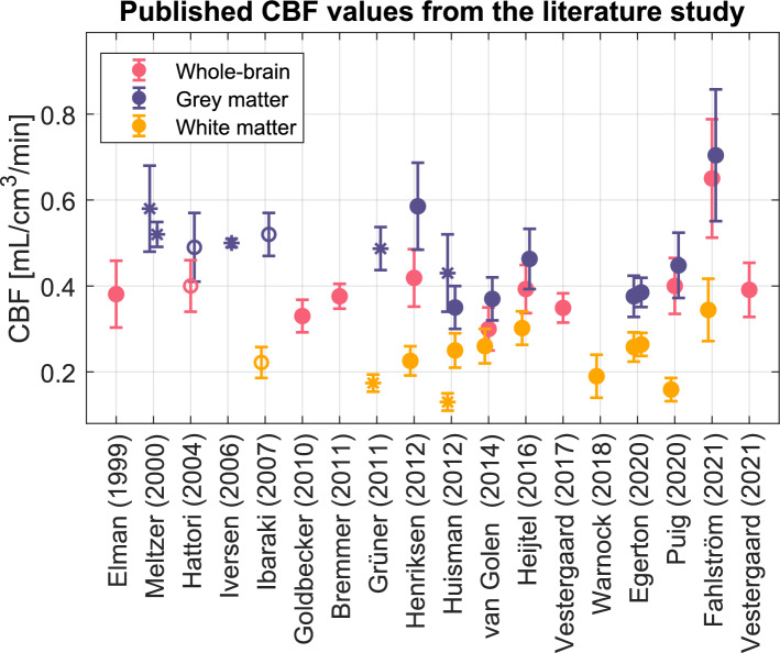
Background: 15O-water positron emission tomography (PET) is considered the gold standard method for non-invasive measurement of cerebral blood flow (CBF). However, previously published average CBF values in healthy subjects have varied greatly and the cause of these variations remains unclear. This study investigates how image reconstruction methods and spatial resolution affect CBF measurements with 15O-water PET.
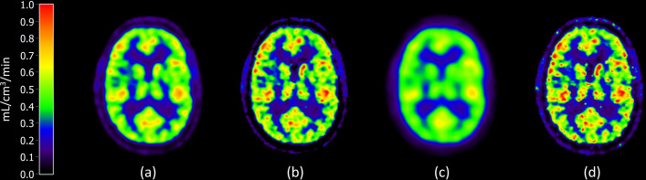
Methods: Eight healthy subjects each underwent dynamic 15O-water PET scans with continuous arterial blood sampling. Images were reconstructed using two different algorithms; ordered subset expectation maximisation and block sequential regularised expectation maximalisation with varying reconstruction parameters. CBF was estimated for the whole brain, grey matter, and central white matter. Reconstruction-specific effective spatial resolution was estimated using phantom measurements and simulations.
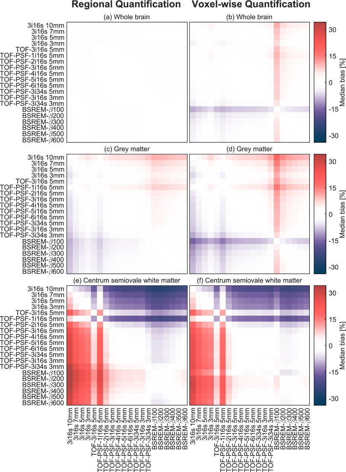
Results: The mean whole brain CBF was 0.48 mL/cm3/min and showed little dependence on the image reconstruction method. Grey matter CBF varied between 0.52 and 0.57 mL/cm3/min, and central white matter CBF between 0.20 and 0.28 mL/cm3/min. Regional CBF showed great dependence on effective spatial resolution with a negative correlation between grey matter CBF and resolution (r = -0.96) and a positive correlation between central white matter and resolution (r = 0.93).
Conclusion: This study concludes that grey matter and central white matter CBF, but not whole brain CBF measured with quantitative 15O-water PET is reconstruction method dependent, mainly due to varying spatial resolution with consequent partial volume effects. Variations in published CBF values cannot be explained solely by reconstruction methods or spatial resolution.
期刊介绍:
EJNMMI Physics is an international platform for scientists, users and adopters of nuclear medicine with a particular interest in physics matters. As a companion journal to the European Journal of Nuclear Medicine and Molecular Imaging, this journal has a multi-disciplinary approach and welcomes original materials and studies with a focus on applied physics and mathematics as well as imaging systems engineering and prototyping in nuclear medicine. This includes physics-driven approaches or algorithms supported by physics that foster early clinical adoption of nuclear medicine imaging and therapy.

 求助内容:
求助内容: 应助结果提醒方式:


