Adrian Jun Zounek, Nico Maximilian Joerg, Felix Lindheimer, Artem Zatcepin, Giovanna Palumbo, Rosel Oos, Astrid Delker, Franz Josef Gildehaus, Andreas Bollenbacher, Guido Boening, Peter Bartenstein, Matthias Brendel, Nathalie Lisa Albert, Sibylle Ziegler, Lena Kaiser
{"title":"3D打印放射性无壁PET幻影改善了基于阈值的目标描绘和量化。","authors":"Adrian Jun Zounek, Nico Maximilian Joerg, Felix Lindheimer, Artem Zatcepin, Giovanna Palumbo, Rosel Oos, Astrid Delker, Franz Josef Gildehaus, Andreas Bollenbacher, Guido Boening, Peter Bartenstein, Matthias Brendel, Nathalie Lisa Albert, Sibylle Ziegler, Lena Kaiser","doi":"10.1186/s40658-025-00768-x","DOIUrl":null,"url":null,"abstract":"<p><strong>Background: </strong>Validation of threshold-based PET segmentation and PET quantification is typically performed with fillable phantoms. Theoretical considerations show that the inactive walls of the phantom cavities introduce a contrast dependence of the volume-reproducing threshold (VRT), potentially leading to segmentation errors and therefore miscalculations of target volumes. The goal of this study was to experimentally show the contrast independence of the VRT when using wall-less phantoms.</p><p><strong>Results: </strong>Radioactive spheres were produced according to NEMA specifications (D = 10/13/17/22/28/37 mm) using a stereolithographic (SLA) 3D printer. For comparison, hollow spheres were filled with a similar activity concentration. Image data from both sphere types were acquired with five different signal-to-background ratios (SBR = 2/4/6/8/10) using a Siemens mCT 20 and a Biograph 64 TruePoint PET/CT system. Results from wall-less and fillable spheres were compared to evaluate contrast dependence and segmentation accuracy based on VRT and intensity profiles. Wall-less phantoms demonstrated consistent VRT values, with a coefficient of variation of 2% over all SBRs, indicating independence from contrast. Conversely, fillable phantoms exhibited significant VRT variability, with a coefficient of variation (CV) of 9% over all SBRs and up to 40% volume overestimation at low contrast. Additionally, activity distribution in the printed spheres was evaluated using PET-based statistical analysis and autoradiography. The PET intensity distribution in the printed material was highly uniform (CV = 4.2%), with a Kullback-Leibler divergence near zero and no statistically significant difference to the fillable spheres. Autoradiography revealed microscopic regions with elevated counts, showing a CV of 11.7%, which was effectively reduced to 2.4% after Gaussian filtering.</p><p><strong>Conclusions: </strong>The theoretical predictions of a significant influence of inactive walls in low-contrast images and contrast-independent VRT in wall-less phantoms were successfully confirmed. SLA 3D printing of phantoms is a promising method for the reliable evaluation of PET quantification methods, particularly in low-contrast scenarios commonly encountered in clinical settings.</p>","PeriodicalId":11559,"journal":{"name":"EJNMMI Physics","volume":"12 1","pages":"53"},"PeriodicalIF":3.2000,"publicationDate":"2025-06-06","publicationTypes":"Journal Article","fieldsOfStudy":null,"isOpenAccess":false,"openAccessPdf":"https://www.ncbi.nlm.nih.gov/pmc/articles/PMC12144006/pdf/","citationCount":"0","resultStr":"{\"title\":\"3D printing of radioactive wall-less PET phantoms improves threshold-based target delineation and quantification.\",\"authors\":\"Adrian Jun Zounek, Nico Maximilian Joerg, Felix Lindheimer, Artem Zatcepin, Giovanna Palumbo, Rosel Oos, Astrid Delker, Franz Josef Gildehaus, Andreas Bollenbacher, Guido Boening, Peter Bartenstein, Matthias Brendel, Nathalie Lisa Albert, Sibylle Ziegler, Lena Kaiser\",\"doi\":\"10.1186/s40658-025-00768-x\",\"DOIUrl\":null,\"url\":null,\"abstract\":\"<p><strong>Background: </strong>Validation of threshold-based PET segmentation and PET quantification is typically performed with fillable phantoms. Theoretical considerations show that the inactive walls of the phantom cavities introduce a contrast dependence of the volume-reproducing threshold (VRT), potentially leading to segmentation errors and therefore miscalculations of target volumes. The goal of this study was to experimentally show the contrast independence of the VRT when using wall-less phantoms.</p><p><strong>Results: </strong>Radioactive spheres were produced according to NEMA specifications (D = 10/13/17/22/28/37 mm) using a stereolithographic (SLA) 3D printer. For comparison, hollow spheres were filled with a similar activity concentration. Image data from both sphere types were acquired with five different signal-to-background ratios (SBR = 2/4/6/8/10) using a Siemens mCT 20 and a Biograph 64 TruePoint PET/CT system. Results from wall-less and fillable spheres were compared to evaluate contrast dependence and segmentation accuracy based on VRT and intensity profiles. Wall-less phantoms demonstrated consistent VRT values, with a coefficient of variation of 2% over all SBRs, indicating independence from contrast. Conversely, fillable phantoms exhibited significant VRT variability, with a coefficient of variation (CV) of 9% over all SBRs and up to 40% volume overestimation at low contrast. Additionally, activity distribution in the printed spheres was evaluated using PET-based statistical analysis and autoradiography. The PET intensity distribution in the printed material was highly uniform (CV = 4.2%), with a Kullback-Leibler divergence near zero and no statistically significant difference to the fillable spheres. Autoradiography revealed microscopic regions with elevated counts, showing a CV of 11.7%, which was effectively reduced to 2.4% after Gaussian filtering.</p><p><strong>Conclusions: </strong>The theoretical predictions of a significant influence of inactive walls in low-contrast images and contrast-independent VRT in wall-less phantoms were successfully confirmed. SLA 3D printing of phantoms is a promising method for the reliable evaluation of PET quantification methods, particularly in low-contrast scenarios commonly encountered in clinical settings.</p>\",\"PeriodicalId\":11559,\"journal\":{\"name\":\"EJNMMI Physics\",\"volume\":\"12 1\",\"pages\":\"53\"},\"PeriodicalIF\":3.2000,\"publicationDate\":\"2025-06-06\",\"publicationTypes\":\"Journal Article\",\"fieldsOfStudy\":null,\"isOpenAccess\":false,\"openAccessPdf\":\"https://www.ncbi.nlm.nih.gov/pmc/articles/PMC12144006/pdf/\",\"citationCount\":\"0\",\"resultStr\":null,\"platform\":\"Semanticscholar\",\"paperid\":null,\"PeriodicalName\":\"EJNMMI Physics\",\"FirstCategoryId\":\"3\",\"ListUrlMain\":\"https://doi.org/10.1186/s40658-025-00768-x\",\"RegionNum\":2,\"RegionCategory\":\"医学\",\"ArticlePicture\":[],\"TitleCN\":null,\"AbstractTextCN\":null,\"PMCID\":null,\"EPubDate\":\"\",\"PubModel\":\"\",\"JCR\":\"Q2\",\"JCRName\":\"RADIOLOGY, NUCLEAR MEDICINE & MEDICAL IMAGING\",\"Score\":null,\"Total\":0}","platform":"Semanticscholar","paperid":null,"PeriodicalName":"EJNMMI Physics","FirstCategoryId":"3","ListUrlMain":"https://doi.org/10.1186/s40658-025-00768-x","RegionNum":2,"RegionCategory":"医学","ArticlePicture":[],"TitleCN":null,"AbstractTextCN":null,"PMCID":null,"EPubDate":"","PubModel":"","JCR":"Q2","JCRName":"RADIOLOGY, NUCLEAR MEDICINE & MEDICAL IMAGING","Score":null,"Total":0}
3D printing of radioactive wall-less PET phantoms improves threshold-based target delineation and quantification.
Background: Validation of threshold-based PET segmentation and PET quantification is typically performed with fillable phantoms. Theoretical considerations show that the inactive walls of the phantom cavities introduce a contrast dependence of the volume-reproducing threshold (VRT), potentially leading to segmentation errors and therefore miscalculations of target volumes. The goal of this study was to experimentally show the contrast independence of the VRT when using wall-less phantoms.
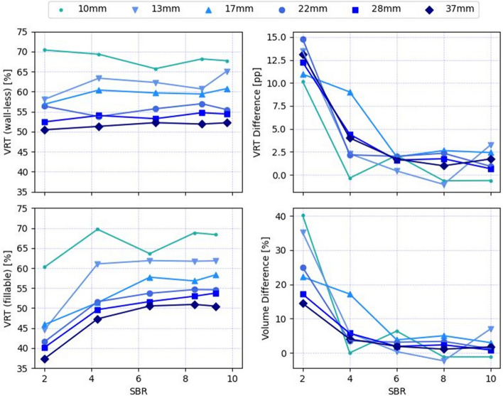
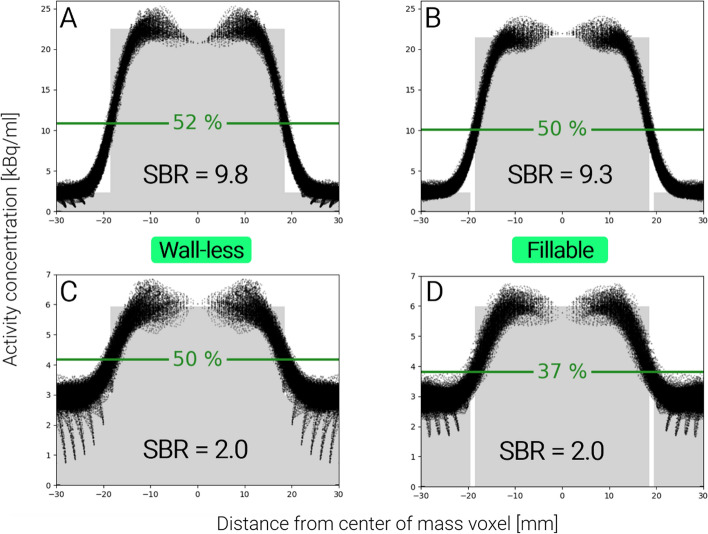
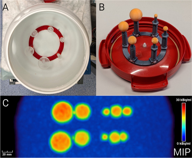
Results: Radioactive spheres were produced according to NEMA specifications (D = 10/13/17/22/28/37 mm) using a stereolithographic (SLA) 3D printer. For comparison, hollow spheres were filled with a similar activity concentration. Image data from both sphere types were acquired with five different signal-to-background ratios (SBR = 2/4/6/8/10) using a Siemens mCT 20 and a Biograph 64 TruePoint PET/CT system. Results from wall-less and fillable spheres were compared to evaluate contrast dependence and segmentation accuracy based on VRT and intensity profiles. Wall-less phantoms demonstrated consistent VRT values, with a coefficient of variation of 2% over all SBRs, indicating independence from contrast. Conversely, fillable phantoms exhibited significant VRT variability, with a coefficient of variation (CV) of 9% over all SBRs and up to 40% volume overestimation at low contrast. Additionally, activity distribution in the printed spheres was evaluated using PET-based statistical analysis and autoradiography. The PET intensity distribution in the printed material was highly uniform (CV = 4.2%), with a Kullback-Leibler divergence near zero and no statistically significant difference to the fillable spheres. Autoradiography revealed microscopic regions with elevated counts, showing a CV of 11.7%, which was effectively reduced to 2.4% after Gaussian filtering.
Conclusions: The theoretical predictions of a significant influence of inactive walls in low-contrast images and contrast-independent VRT in wall-less phantoms were successfully confirmed. SLA 3D printing of phantoms is a promising method for the reliable evaluation of PET quantification methods, particularly in low-contrast scenarios commonly encountered in clinical settings.
期刊介绍:
EJNMMI Physics is an international platform for scientists, users and adopters of nuclear medicine with a particular interest in physics matters. As a companion journal to the European Journal of Nuclear Medicine and Molecular Imaging, this journal has a multi-disciplinary approach and welcomes original materials and studies with a focus on applied physics and mathematics as well as imaging systems engineering and prototyping in nuclear medicine. This includes physics-driven approaches or algorithms supported by physics that foster early clinical adoption of nuclear medicine imaging and therapy.

 求助内容:
求助内容: 应助结果提醒方式:


