Tilman A. Grünewald, Peng Li, Julien Duboisset, Julius Nouet, Oier Bikondoa, Jeremie Vidal-Dupiol, Denis Saulnier, Manfred Burghammer and Virginie Chamard
{"title":"三维Bragg图谱研究生物矿物软体动物壳的结晶。","authors":"Tilman A. Grünewald, Peng Li, Julien Duboisset, Julius Nouet, Oier Bikondoa, Jeremie Vidal-Dupiol, Denis Saulnier, Manfred Burghammer and Virginie Chamard","doi":"10.1039/D5FD00020C","DOIUrl":null,"url":null,"abstract":"<p >Biomineralisation integrates complex biologically assisted physico-chemical processes leading to an extraordinary diversity of calcareous biomineral crystalline architectures, in intriguing contrast with the consistent presence of a submicrometric granular structure. While the repeated observation of amorphous calcium carbonate is interpreted as a precursor to the crystalline phase, the crystalline transition mechanisms are poorly understood. Access to the crystalline architecture at the mesoscale, <em>i.e.</em>, over a few granules, is key to building realistic crystallisation models. Here we exploit three-dimensional X-ray Bragg ptychography microscopy to provide a series of nanoscale maps of the crystalline structure within the “single-crystalline” prism of the prismatic layer of a <em>Pinctada margaritifera</em> shell. The mesocrystalline organisation exhibits several micrometre-sized iso-oriented/iso-strained crystalline domains, the detailed studies of which reveal the presence of crystalline coherence domains ranging from 130 to 550 nm in size. The further increase in the lattice parameter with the size of the coherence domain likely results from the crystallisation mechanism, pointing towards a maturation process occurring after the initial amorphous-to-crystalline transition.</p>","PeriodicalId":49075,"journal":{"name":"Faraday Discussions","volume":"261 ","pages":" 192-211"},"PeriodicalIF":3.1000,"publicationDate":"2025-06-06","publicationTypes":"Journal Article","fieldsOfStudy":null,"isOpenAccess":false,"openAccessPdf":"","citationCount":"0","resultStr":"{\"title\":\"Crystallisation in biomineral mollusc shell studied by 3D Bragg ptychography\",\"authors\":\"Tilman A. Grünewald, Peng Li, Julien Duboisset, Julius Nouet, Oier Bikondoa, Jeremie Vidal-Dupiol, Denis Saulnier, Manfred Burghammer and Virginie Chamard\",\"doi\":\"10.1039/D5FD00020C\",\"DOIUrl\":null,\"url\":null,\"abstract\":\"<p >Biomineralisation integrates complex biologically assisted physico-chemical processes leading to an extraordinary diversity of calcareous biomineral crystalline architectures, in intriguing contrast with the consistent presence of a submicrometric granular structure. While the repeated observation of amorphous calcium carbonate is interpreted as a precursor to the crystalline phase, the crystalline transition mechanisms are poorly understood. Access to the crystalline architecture at the mesoscale, <em>i.e.</em>, over a few granules, is key to building realistic crystallisation models. Here we exploit three-dimensional X-ray Bragg ptychography microscopy to provide a series of nanoscale maps of the crystalline structure within the “single-crystalline” prism of the prismatic layer of a <em>Pinctada margaritifera</em> shell. The mesocrystalline organisation exhibits several micrometre-sized iso-oriented/iso-strained crystalline domains, the detailed studies of which reveal the presence of crystalline coherence domains ranging from 130 to 550 nm in size. The further increase in the lattice parameter with the size of the coherence domain likely results from the crystallisation mechanism, pointing towards a maturation process occurring after the initial amorphous-to-crystalline transition.</p>\",\"PeriodicalId\":49075,\"journal\":{\"name\":\"Faraday Discussions\",\"volume\":\"261 \",\"pages\":\" 192-211\"},\"PeriodicalIF\":3.1000,\"publicationDate\":\"2025-06-06\",\"publicationTypes\":\"Journal Article\",\"fieldsOfStudy\":null,\"isOpenAccess\":false,\"openAccessPdf\":\"\",\"citationCount\":\"0\",\"resultStr\":null,\"platform\":\"Semanticscholar\",\"paperid\":null,\"PeriodicalName\":\"Faraday Discussions\",\"FirstCategoryId\":\"92\",\"ListUrlMain\":\"https://pubs.rsc.org/en/content/articlelanding/2025/fd/d5fd00020c\",\"RegionNum\":3,\"RegionCategory\":\"化学\",\"ArticlePicture\":[],\"TitleCN\":null,\"AbstractTextCN\":null,\"PMCID\":null,\"EPubDate\":\"\",\"PubModel\":\"\",\"JCR\":\"Q2\",\"JCRName\":\"Chemistry\",\"Score\":null,\"Total\":0}","platform":"Semanticscholar","paperid":null,"PeriodicalName":"Faraday Discussions","FirstCategoryId":"92","ListUrlMain":"https://pubs.rsc.org/en/content/articlelanding/2025/fd/d5fd00020c","RegionNum":3,"RegionCategory":"化学","ArticlePicture":[],"TitleCN":null,"AbstractTextCN":null,"PMCID":null,"EPubDate":"","PubModel":"","JCR":"Q2","JCRName":"Chemistry","Score":null,"Total":0}
Crystallisation in biomineral mollusc shell studied by 3D Bragg ptychography
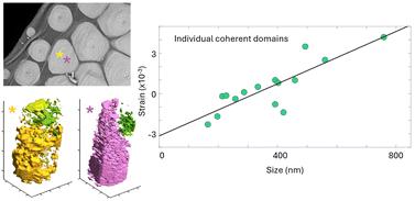
Biomineralisation integrates complex biologically assisted physico-chemical processes leading to an extraordinary diversity of calcareous biomineral crystalline architectures, in intriguing contrast with the consistent presence of a submicrometric granular structure. While the repeated observation of amorphous calcium carbonate is interpreted as a precursor to the crystalline phase, the crystalline transition mechanisms are poorly understood. Access to the crystalline architecture at the mesoscale, i.e., over a few granules, is key to building realistic crystallisation models. Here we exploit three-dimensional X-ray Bragg ptychography microscopy to provide a series of nanoscale maps of the crystalline structure within the “single-crystalline” prism of the prismatic layer of a Pinctada margaritifera shell. The mesocrystalline organisation exhibits several micrometre-sized iso-oriented/iso-strained crystalline domains, the detailed studies of which reveal the presence of crystalline coherence domains ranging from 130 to 550 nm in size. The further increase in the lattice parameter with the size of the coherence domain likely results from the crystallisation mechanism, pointing towards a maturation process occurring after the initial amorphous-to-crystalline transition.
 求助内容:
求助内容: 应助结果提醒方式:


