Musawenkosi M Mthombeni, Nasreen Mahomed, Grace Rubin, Sharadini K Gounden
{"title":"乳头状乳腺癌的妇女出席乳房成像中心在约翰内斯堡的回顾。","authors":"Musawenkosi M Mthombeni, Nasreen Mahomed, Grace Rubin, Sharadini K Gounden","doi":"10.4102/sajr.v29i1.3092","DOIUrl":null,"url":null,"abstract":"<p><strong>Background: </strong>Breast cancer ranks globally as the most prevalent cause of female deaths. Papillary breast carcinoma (PBC), a rare subtype of breast cancer, presents distinct challenges in diagnosis and management because of its unique histopathological features.</p><p><strong>Objectives: </strong>This study aims to determine the prevalence and main imaging findings of PBC in women attending a tertiary breast imaging centre.</p><p><strong>Method: </strong>A retrospective review of mammography and ultrasound imaging findings of female patients with histologically proven PBC, referred to a tertiary breast imaging centre over a 5-year period, was conducted.</p><p><strong>Results: </strong>The study included 102 female patients with a mean age of 53.8. Mammography detected masses in 93.02%, with calcifications in 41.2% and abnormal borders in 56.8%. Architectural distortion and asymmetry occurred in 27.5% and 28.4% respectively, both showing moderate correlation with PBC (<i>r</i> = 0.50, <i>p</i> = 0.009; <i>r</i> = 0.51, <i>p</i> = 0.0057). Ultrasound findings indicated irregular mass shapes (mean = 1.53), with hypoechoic patterns significantly associated with PBC (<i>r</i> = 0.40, <i>p</i> = 0.0013). Correlation analysis revealed strong associations between PBC and breast pain (<i>r</i> = 0.74, <i>p</i> < 0.0001), and erythema (<i>r</i> = 0.62, <i>p</i> < 0.0001). There was no significant association between the mammography and ultrasound findings (<i>p</i> = 0.495).</p><p><strong>Conclusion: </strong>The findings underscore the value of using mammography and ultrasound in the diagnosis of PBC, as the two modalities offer complementary information.</p><p><strong>Contribution: </strong>There is a paucity of data on the radiological findings of PBC in Africa. The current study prevalence mirrors global trends, highlighting the importance of ongoing surveillance and diagnostic accuracy.</p>","PeriodicalId":43442,"journal":{"name":"SA Journal of Radiology","volume":"29 1","pages":"3092"},"PeriodicalIF":0.9000,"publicationDate":"2025-05-02","publicationTypes":"Journal Article","fieldsOfStudy":null,"isOpenAccess":false,"openAccessPdf":"https://www.ncbi.nlm.nih.gov/pmc/articles/PMC12135729/pdf/","citationCount":"0","resultStr":"{\"title\":\"A review of papillary breast carcinoma in women attending a breast imaging centre in Johannesburg.\",\"authors\":\"Musawenkosi M Mthombeni, Nasreen Mahomed, Grace Rubin, Sharadini K Gounden\",\"doi\":\"10.4102/sajr.v29i1.3092\",\"DOIUrl\":null,\"url\":null,\"abstract\":\"<p><strong>Background: </strong>Breast cancer ranks globally as the most prevalent cause of female deaths. Papillary breast carcinoma (PBC), a rare subtype of breast cancer, presents distinct challenges in diagnosis and management because of its unique histopathological features.</p><p><strong>Objectives: </strong>This study aims to determine the prevalence and main imaging findings of PBC in women attending a tertiary breast imaging centre.</p><p><strong>Method: </strong>A retrospective review of mammography and ultrasound imaging findings of female patients with histologically proven PBC, referred to a tertiary breast imaging centre over a 5-year period, was conducted.</p><p><strong>Results: </strong>The study included 102 female patients with a mean age of 53.8. Mammography detected masses in 93.02%, with calcifications in 41.2% and abnormal borders in 56.8%. Architectural distortion and asymmetry occurred in 27.5% and 28.4% respectively, both showing moderate correlation with PBC (<i>r</i> = 0.50, <i>p</i> = 0.009; <i>r</i> = 0.51, <i>p</i> = 0.0057). Ultrasound findings indicated irregular mass shapes (mean = 1.53), with hypoechoic patterns significantly associated with PBC (<i>r</i> = 0.40, <i>p</i> = 0.0013). Correlation analysis revealed strong associations between PBC and breast pain (<i>r</i> = 0.74, <i>p</i> < 0.0001), and erythema (<i>r</i> = 0.62, <i>p</i> < 0.0001). There was no significant association between the mammography and ultrasound findings (<i>p</i> = 0.495).</p><p><strong>Conclusion: </strong>The findings underscore the value of using mammography and ultrasound in the diagnosis of PBC, as the two modalities offer complementary information.</p><p><strong>Contribution: </strong>There is a paucity of data on the radiological findings of PBC in Africa. The current study prevalence mirrors global trends, highlighting the importance of ongoing surveillance and diagnostic accuracy.</p>\",\"PeriodicalId\":43442,\"journal\":{\"name\":\"SA Journal of Radiology\",\"volume\":\"29 1\",\"pages\":\"3092\"},\"PeriodicalIF\":0.9000,\"publicationDate\":\"2025-05-02\",\"publicationTypes\":\"Journal Article\",\"fieldsOfStudy\":null,\"isOpenAccess\":false,\"openAccessPdf\":\"https://www.ncbi.nlm.nih.gov/pmc/articles/PMC12135729/pdf/\",\"citationCount\":\"0\",\"resultStr\":null,\"platform\":\"Semanticscholar\",\"paperid\":null,\"PeriodicalName\":\"SA Journal of Radiology\",\"FirstCategoryId\":\"1085\",\"ListUrlMain\":\"https://doi.org/10.4102/sajr.v29i1.3092\",\"RegionNum\":0,\"RegionCategory\":null,\"ArticlePicture\":[],\"TitleCN\":null,\"AbstractTextCN\":null,\"PMCID\":null,\"EPubDate\":\"2025/1/1 0:00:00\",\"PubModel\":\"eCollection\",\"JCR\":\"Q4\",\"JCRName\":\"RADIOLOGY, NUCLEAR MEDICINE & MEDICAL IMAGING\",\"Score\":null,\"Total\":0}","platform":"Semanticscholar","paperid":null,"PeriodicalName":"SA Journal of Radiology","FirstCategoryId":"1085","ListUrlMain":"https://doi.org/10.4102/sajr.v29i1.3092","RegionNum":0,"RegionCategory":null,"ArticlePicture":[],"TitleCN":null,"AbstractTextCN":null,"PMCID":null,"EPubDate":"2025/1/1 0:00:00","PubModel":"eCollection","JCR":"Q4","JCRName":"RADIOLOGY, NUCLEAR MEDICINE & MEDICAL IMAGING","Score":null,"Total":0}
引用次数: 0
摘要
背景:乳腺癌在全球范围内是导致女性死亡的最普遍原因。乳头状乳腺癌(PBC)是一种罕见的乳腺癌亚型,由于其独特的组织病理学特征,在诊断和治疗方面面临着明显的挑战。目的:本研究旨在确定在三级乳腺成像中心就诊的女性中PBC的患病率和主要影像学表现。方法:回顾性分析经组织学证实的PBC女性患者的乳腺x线摄影和超声成像结果,并在三级乳腺成像中心进行了5年的研究。结果:纳入102例女性患者,平均年龄53.8岁。乳腺x线检查发现肿块93.02%,钙化41.2%,边界异常56.8%。建筑变形和不对称发生率分别为27.5%和28.4%,两者均与PBC呈中度相关(r = 0.50, p = 0.009;R = 0.51, p = 0.0057)。超声结果显示不规则肿块形状(平均= 1.53),低回声模式与PBC显著相关(r = 0.40, p = 0.0013)。相关分析显示PBC与乳房疼痛(r = 0.74, p < 0.0001)和红斑(r = 0.62, p < 0.0001)有很强的相关性。乳腺x光检查与超声检查结果无显著相关性(p = 0.495)。结论:这些发现强调了使用乳房x线摄影和超声诊断PBC的价值,因为这两种方式提供了互补的信息。贡献:非洲PBC的放射学表现数据缺乏。目前的研究流行率反映了全球趋势,突出了持续监测和诊断准确性的重要性。
A review of papillary breast carcinoma in women attending a breast imaging centre in Johannesburg.
Background: Breast cancer ranks globally as the most prevalent cause of female deaths. Papillary breast carcinoma (PBC), a rare subtype of breast cancer, presents distinct challenges in diagnosis and management because of its unique histopathological features.
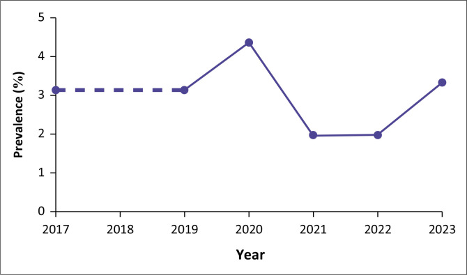
Objectives: This study aims to determine the prevalence and main imaging findings of PBC in women attending a tertiary breast imaging centre.
Method: A retrospective review of mammography and ultrasound imaging findings of female patients with histologically proven PBC, referred to a tertiary breast imaging centre over a 5-year period, was conducted.
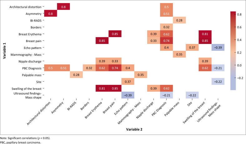
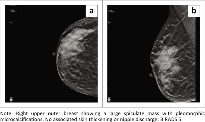
Results: The study included 102 female patients with a mean age of 53.8. Mammography detected masses in 93.02%, with calcifications in 41.2% and abnormal borders in 56.8%. Architectural distortion and asymmetry occurred in 27.5% and 28.4% respectively, both showing moderate correlation with PBC (r = 0.50, p = 0.009; r = 0.51, p = 0.0057). Ultrasound findings indicated irregular mass shapes (mean = 1.53), with hypoechoic patterns significantly associated with PBC (r = 0.40, p = 0.0013). Correlation analysis revealed strong associations between PBC and breast pain (r = 0.74, p < 0.0001), and erythema (r = 0.62, p < 0.0001). There was no significant association between the mammography and ultrasound findings (p = 0.495).
Conclusion: The findings underscore the value of using mammography and ultrasound in the diagnosis of PBC, as the two modalities offer complementary information.
Contribution: There is a paucity of data on the radiological findings of PBC in Africa. The current study prevalence mirrors global trends, highlighting the importance of ongoing surveillance and diagnostic accuracy.
期刊介绍:
The SA Journal of Radiology is the official journal of the Radiological Society of South Africa and the Professional Association of Radiologists in South Africa and Namibia. The SA Journal of Radiology is a general diagnostic radiological journal which carries original research and review articles, pictorial essays, case reports, letters, editorials, radiological practice and other radiological articles.

 求助内容:
求助内容: 应助结果提醒方式:


