Lexi Gower-Fry, Justin J. Bailey, Melinda Wuest, Susan Pike, Alexey Kostikov, Andreas Dorian, Carmen Wängler, Frank Wuest and Ralf Schirrmacher
{"title":"18F-PSiMA在前列腺癌PET成像中的发展及临床潜力。","authors":"Lexi Gower-Fry, Justin J. Bailey, Melinda Wuest, Susan Pike, Alexey Kostikov, Andreas Dorian, Carmen Wängler, Frank Wuest and Ralf Schirrmacher","doi":"10.1039/D5MD00275C","DOIUrl":null,"url":null,"abstract":"<p >Prostate-specific membrane antigen (PSMA) is a key target for diagnosing prostate cancer through positron emission tomography (PET). While <small><sup>68</sup></small>Ga-labeled PSMA compounds are widely used, <small><sup>18</sup></small>F-labeled PSMA inhibitors have gained traction for clinical tumor imaging. We previously investigated PSMA-targeting compounds based on the Lys-urea-Glu motif, incorporating a silicon fluoride-acceptor (SiFA) and chemical auxiliaries to enhance <em>in vivo</em> biodistribution. This led to the development of <small><sup>18</sup></small>F-PSiMA, a SiFA-based radiotracer with an optimized linker exhibiting favorable PSMA potency (IC<small><sub>50</sub></small> = 154 ± 47 nM in LNCaP cells). <small><sup>18</sup></small>F-PSiMA radiosynthesis with low to high concentrations of <small><sup>18</sup></small>F and precursor achieved molar activities (<em>A</em><small><sub>m</sub></small>) of 10.9–82.5 GBq μmol<small><sup>−1</sup></small> and showed a 24–38% increase in tumor uptake in LNCaP tumors (SUV<small><sub>60min</sub></small> 1.56 ± 0.18; 7.23 ± 0.75% ID per g at lower <em>A</em><small><sub>m</sub></small> and SUV<small><sub>60min</sub></small> 1.90 ± 0.29; 9.62 ± 1.29% ID per g at higher <em>A</em><small><sub>m</sub></small>) compared to our previous lead, <small><sup>18</sup></small>F-SiFA-Asp<small><sub>2</sub></small>-PEG<small><sub>3</sub></small>-PSMA. PSMA specificity was confirmed by a 20 ± 10% reduction in SUV<small><sub>60min</sub></small> upon co-injection with DCFPyl. These promising <em>in vitro</em> and <em>in vivo</em> results support further clinical translation of <small><sup>18</sup></small>F-PSiMA for prostate cancer PET imaging.</p>","PeriodicalId":21462,"journal":{"name":"RSC medicinal chemistry","volume":" 8","pages":" 3633-3644"},"PeriodicalIF":3.6000,"publicationDate":"2025-05-09","publicationTypes":"Journal Article","fieldsOfStudy":null,"isOpenAccess":false,"openAccessPdf":"https://www.ncbi.nlm.nih.gov/pmc/articles/PMC12127850/pdf/","citationCount":"0","resultStr":"{\"title\":\"Development and clinical potential of 18F-PSiMA for prostate cancer PET imaging†\",\"authors\":\"Lexi Gower-Fry, Justin J. Bailey, Melinda Wuest, Susan Pike, Alexey Kostikov, Andreas Dorian, Carmen Wängler, Frank Wuest and Ralf Schirrmacher\",\"doi\":\"10.1039/D5MD00275C\",\"DOIUrl\":null,\"url\":null,\"abstract\":\"<p >Prostate-specific membrane antigen (PSMA) is a key target for diagnosing prostate cancer through positron emission tomography (PET). While <small><sup>68</sup></small>Ga-labeled PSMA compounds are widely used, <small><sup>18</sup></small>F-labeled PSMA inhibitors have gained traction for clinical tumor imaging. We previously investigated PSMA-targeting compounds based on the Lys-urea-Glu motif, incorporating a silicon fluoride-acceptor (SiFA) and chemical auxiliaries to enhance <em>in vivo</em> biodistribution. This led to the development of <small><sup>18</sup></small>F-PSiMA, a SiFA-based radiotracer with an optimized linker exhibiting favorable PSMA potency (IC<small><sub>50</sub></small> = 154 ± 47 nM in LNCaP cells). <small><sup>18</sup></small>F-PSiMA radiosynthesis with low to high concentrations of <small><sup>18</sup></small>F and precursor achieved molar activities (<em>A</em><small><sub>m</sub></small>) of 10.9–82.5 GBq μmol<small><sup>−1</sup></small> and showed a 24–38% increase in tumor uptake in LNCaP tumors (SUV<small><sub>60min</sub></small> 1.56 ± 0.18; 7.23 ± 0.75% ID per g at lower <em>A</em><small><sub>m</sub></small> and SUV<small><sub>60min</sub></small> 1.90 ± 0.29; 9.62 ± 1.29% ID per g at higher <em>A</em><small><sub>m</sub></small>) compared to our previous lead, <small><sup>18</sup></small>F-SiFA-Asp<small><sub>2</sub></small>-PEG<small><sub>3</sub></small>-PSMA. PSMA specificity was confirmed by a 20 ± 10% reduction in SUV<small><sub>60min</sub></small> upon co-injection with DCFPyl. These promising <em>in vitro</em> and <em>in vivo</em> results support further clinical translation of <small><sup>18</sup></small>F-PSiMA for prostate cancer PET imaging.</p>\",\"PeriodicalId\":21462,\"journal\":{\"name\":\"RSC medicinal chemistry\",\"volume\":\" 8\",\"pages\":\" 3633-3644\"},\"PeriodicalIF\":3.6000,\"publicationDate\":\"2025-05-09\",\"publicationTypes\":\"Journal Article\",\"fieldsOfStudy\":null,\"isOpenAccess\":false,\"openAccessPdf\":\"https://www.ncbi.nlm.nih.gov/pmc/articles/PMC12127850/pdf/\",\"citationCount\":\"0\",\"resultStr\":null,\"platform\":\"Semanticscholar\",\"paperid\":null,\"PeriodicalName\":\"RSC medicinal chemistry\",\"FirstCategoryId\":\"3\",\"ListUrlMain\":\"https://pubs.rsc.org/en/content/articlelanding/2025/md/d5md00275c\",\"RegionNum\":4,\"RegionCategory\":\"医学\",\"ArticlePicture\":[],\"TitleCN\":null,\"AbstractTextCN\":null,\"PMCID\":null,\"EPubDate\":\"\",\"PubModel\":\"\",\"JCR\":\"Q2\",\"JCRName\":\"BIOCHEMISTRY & MOLECULAR BIOLOGY\",\"Score\":null,\"Total\":0}","platform":"Semanticscholar","paperid":null,"PeriodicalName":"RSC medicinal chemistry","FirstCategoryId":"3","ListUrlMain":"https://pubs.rsc.org/en/content/articlelanding/2025/md/d5md00275c","RegionNum":4,"RegionCategory":"医学","ArticlePicture":[],"TitleCN":null,"AbstractTextCN":null,"PMCID":null,"EPubDate":"","PubModel":"","JCR":"Q2","JCRName":"BIOCHEMISTRY & MOLECULAR BIOLOGY","Score":null,"Total":0}
Development and clinical potential of 18F-PSiMA for prostate cancer PET imaging†
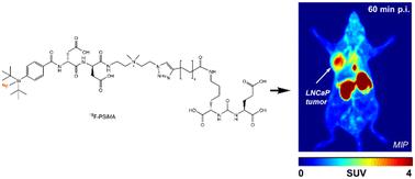
Prostate-specific membrane antigen (PSMA) is a key target for diagnosing prostate cancer through positron emission tomography (PET). While 68Ga-labeled PSMA compounds are widely used, 18F-labeled PSMA inhibitors have gained traction for clinical tumor imaging. We previously investigated PSMA-targeting compounds based on the Lys-urea-Glu motif, incorporating a silicon fluoride-acceptor (SiFA) and chemical auxiliaries to enhance in vivo biodistribution. This led to the development of 18F-PSiMA, a SiFA-based radiotracer with an optimized linker exhibiting favorable PSMA potency (IC50 = 154 ± 47 nM in LNCaP cells). 18F-PSiMA radiosynthesis with low to high concentrations of 18F and precursor achieved molar activities (Am) of 10.9–82.5 GBq μmol−1 and showed a 24–38% increase in tumor uptake in LNCaP tumors (SUV60min 1.56 ± 0.18; 7.23 ± 0.75% ID per g at lower Am and SUV60min 1.90 ± 0.29; 9.62 ± 1.29% ID per g at higher Am) compared to our previous lead, 18F-SiFA-Asp2-PEG3-PSMA. PSMA specificity was confirmed by a 20 ± 10% reduction in SUV60min upon co-injection with DCFPyl. These promising in vitro and in vivo results support further clinical translation of 18F-PSiMA for prostate cancer PET imaging.
 求助内容:
求助内容: 应助结果提醒方式:


