Razan Yasser Alauti, Areej Hussein Mukhtar, Mohammed Alasqah, Mohammed H Alenazi, Khalid Gufran
{"title":"不同浓度褪黑素对培养人牙周韧带干细胞的影响。","authors":"Razan Yasser Alauti, Areej Hussein Mukhtar, Mohammed Alasqah, Mohammed H Alenazi, Khalid Gufran","doi":"10.12659/MSMBR.948551","DOIUrl":null,"url":null,"abstract":"<p><p>BACKGROUND Due to their good differentiation abilities, human periodontal ligament stem cells (hPDLSCs) and tissue engineering treatment modalities are used to treat bone defects due to periodontitis. This in-vitro study aimed to determine the effects of different melatonin concentrations on the viability of cultured periodontal ligament stem cells. MATERIAL AND METHODS Cultured hPLDSCs were treated with different melatonin concentrations. The study groups were divided into 9 concentration-based groups: Group 1, untreated hPDLSCs; Group 2, hPDLSCs exposed to physiological doses of melatonin (1 μM); Group 3, hPDLSCs exposed to 10 μM melatonin; Group 4, hPDLSCs exposed to 20 μM melatonin; Group 5, hPDLSCs exposed to 30 μM melatonin; Group 6, hPDLSCs exposed to 40 μM melatonin; Group 7, hPDLSCs exposed to 50 μM melatonin; Group 8, hPDLSCs exposed to 100 μM melatonin; Group 9, hPDLSCs exposed to 150 μM melatonin. Cell viability was evaluated at 14 and 21 days using the AlamarBlue (AB) Cell Viability Assay, a toxicity assay. RESULTS Melatonin had a positive effect on hPDLSC viability in all test groups. A general pattern of decreasing cell viability with increasing melatonin concentrations was observed, with Groups 8 and 9 showing the lowest viability. An abrupt pattern was detected in Groups 4 and 7, where a sudden, significant increase in cell viability was observed, in contrast with the general observed pattern. CONCLUSIONS In conclusion, the hPDLSCs remained viable when exposed to the melatonin doses used, under osteogenic media, except for the 100µM and 150 µM melatonin groups. Therefore, further studies are required with hPDLSCs exposed to different concentrations of melatonin.</p>","PeriodicalId":18491,"journal":{"name":"Medical Science Monitor Basic Research","volume":"31 ","pages":"e948551"},"PeriodicalIF":2.0000,"publicationDate":"2025-06-03","publicationTypes":"Journal Article","fieldsOfStudy":null,"isOpenAccess":false,"openAccessPdf":"https://www.ncbi.nlm.nih.gov/pmc/articles/PMC12144917/pdf/","citationCount":"0","resultStr":"{\"title\":\"Efficacy of Different Concentrations of Melatonin on Cultured Human Periodontal Ligament Stem Cells.\",\"authors\":\"Razan Yasser Alauti, Areej Hussein Mukhtar, Mohammed Alasqah, Mohammed H Alenazi, Khalid Gufran\",\"doi\":\"10.12659/MSMBR.948551\",\"DOIUrl\":null,\"url\":null,\"abstract\":\"<p><p>BACKGROUND Due to their good differentiation abilities, human periodontal ligament stem cells (hPDLSCs) and tissue engineering treatment modalities are used to treat bone defects due to periodontitis. This in-vitro study aimed to determine the effects of different melatonin concentrations on the viability of cultured periodontal ligament stem cells. MATERIAL AND METHODS Cultured hPLDSCs were treated with different melatonin concentrations. The study groups were divided into 9 concentration-based groups: Group 1, untreated hPDLSCs; Group 2, hPDLSCs exposed to physiological doses of melatonin (1 μM); Group 3, hPDLSCs exposed to 10 μM melatonin; Group 4, hPDLSCs exposed to 20 μM melatonin; Group 5, hPDLSCs exposed to 30 μM melatonin; Group 6, hPDLSCs exposed to 40 μM melatonin; Group 7, hPDLSCs exposed to 50 μM melatonin; Group 8, hPDLSCs exposed to 100 μM melatonin; Group 9, hPDLSCs exposed to 150 μM melatonin. Cell viability was evaluated at 14 and 21 days using the AlamarBlue (AB) Cell Viability Assay, a toxicity assay. RESULTS Melatonin had a positive effect on hPDLSC viability in all test groups. A general pattern of decreasing cell viability with increasing melatonin concentrations was observed, with Groups 8 and 9 showing the lowest viability. An abrupt pattern was detected in Groups 4 and 7, where a sudden, significant increase in cell viability was observed, in contrast with the general observed pattern. CONCLUSIONS In conclusion, the hPDLSCs remained viable when exposed to the melatonin doses used, under osteogenic media, except for the 100µM and 150 µM melatonin groups. Therefore, further studies are required with hPDLSCs exposed to different concentrations of melatonin.</p>\",\"PeriodicalId\":18491,\"journal\":{\"name\":\"Medical Science Monitor Basic Research\",\"volume\":\"31 \",\"pages\":\"e948551\"},\"PeriodicalIF\":2.0000,\"publicationDate\":\"2025-06-03\",\"publicationTypes\":\"Journal Article\",\"fieldsOfStudy\":null,\"isOpenAccess\":false,\"openAccessPdf\":\"https://www.ncbi.nlm.nih.gov/pmc/articles/PMC12144917/pdf/\",\"citationCount\":\"0\",\"resultStr\":null,\"platform\":\"Semanticscholar\",\"paperid\":null,\"PeriodicalName\":\"Medical Science Monitor Basic Research\",\"FirstCategoryId\":\"1085\",\"ListUrlMain\":\"https://doi.org/10.12659/MSMBR.948551\",\"RegionNum\":0,\"RegionCategory\":null,\"ArticlePicture\":[],\"TitleCN\":null,\"AbstractTextCN\":null,\"PMCID\":null,\"EPubDate\":\"\",\"PubModel\":\"\",\"JCR\":\"Q3\",\"JCRName\":\"MEDICINE, RESEARCH & EXPERIMENTAL\",\"Score\":null,\"Total\":0}","platform":"Semanticscholar","paperid":null,"PeriodicalName":"Medical Science Monitor Basic Research","FirstCategoryId":"1085","ListUrlMain":"https://doi.org/10.12659/MSMBR.948551","RegionNum":0,"RegionCategory":null,"ArticlePicture":[],"TitleCN":null,"AbstractTextCN":null,"PMCID":null,"EPubDate":"","PubModel":"","JCR":"Q3","JCRName":"MEDICINE, RESEARCH & EXPERIMENTAL","Score":null,"Total":0}
Efficacy of Different Concentrations of Melatonin on Cultured Human Periodontal Ligament Stem Cells.
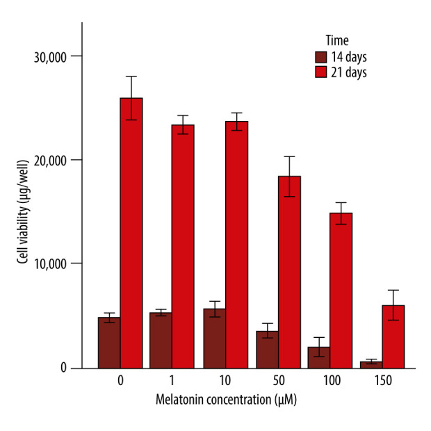
BACKGROUND Due to their good differentiation abilities, human periodontal ligament stem cells (hPDLSCs) and tissue engineering treatment modalities are used to treat bone defects due to periodontitis. This in-vitro study aimed to determine the effects of different melatonin concentrations on the viability of cultured periodontal ligament stem cells. MATERIAL AND METHODS Cultured hPLDSCs were treated with different melatonin concentrations. The study groups were divided into 9 concentration-based groups: Group 1, untreated hPDLSCs; Group 2, hPDLSCs exposed to physiological doses of melatonin (1 μM); Group 3, hPDLSCs exposed to 10 μM melatonin; Group 4, hPDLSCs exposed to 20 μM melatonin; Group 5, hPDLSCs exposed to 30 μM melatonin; Group 6, hPDLSCs exposed to 40 μM melatonin; Group 7, hPDLSCs exposed to 50 μM melatonin; Group 8, hPDLSCs exposed to 100 μM melatonin; Group 9, hPDLSCs exposed to 150 μM melatonin. Cell viability was evaluated at 14 and 21 days using the AlamarBlue (AB) Cell Viability Assay, a toxicity assay. RESULTS Melatonin had a positive effect on hPDLSC viability in all test groups. A general pattern of decreasing cell viability with increasing melatonin concentrations was observed, with Groups 8 and 9 showing the lowest viability. An abrupt pattern was detected in Groups 4 and 7, where a sudden, significant increase in cell viability was observed, in contrast with the general observed pattern. CONCLUSIONS In conclusion, the hPDLSCs remained viable when exposed to the melatonin doses used, under osteogenic media, except for the 100µM and 150 µM melatonin groups. Therefore, further studies are required with hPDLSCs exposed to different concentrations of melatonin.

 求助内容:
求助内容: 应助结果提醒方式:


