基于深度图像先验和一种新的参数放大策略的动态PET直接参数重建

IF 6.3 2区 医学 Q1 BIOLOGY
Xiaotong Hong , Fanghu Wang , Hao Sun , Hossein Arabi , Lijun Lu
{"title":"基于深度图像先验和一种新的参数放大策略的动态PET直接参数重建","authors":"Xiaotong Hong ,&nbsp;Fanghu Wang ,&nbsp;Hao Sun ,&nbsp;Hossein Arabi ,&nbsp;Lijun Lu","doi":"10.1016/j.compbiomed.2025.110487","DOIUrl":null,"url":null,"abstract":"<div><h3>Background/Purpose</h3><div>Multiple parametric imaging in positron emission tomography (PET) is challenging due to the noisy dynamic data and the complex mapping to kinetic parameters. Although methods like direct parametric reconstruction have been proposed to improve the image quality, limitations persist, particularly for nonlinear and small-value micro-parameters (e.g., k<sub>2</sub>, k<sub>3</sub>). This study presents a novel unsupervised deep learning approach to reconstruct and improve the quality of these micro-parameters.</div></div><div><h3>Methods</h3><div>We proposed a direct parametric image reconstruction model, DIP-PM, integrating deep image prior (DIP) with a parameter magnification (PM) strategy. The model employs a U-Net generator to predict multiple parametric images using a CT image prior, with each output channel subsequently magnified by a factor to adjust the intensity. The model was optimized with a log-likelihood loss computed between the measured projection data and forward projected data. Two tracer datasets were simulated for evaluation: <sup>82</sup>Rb data using the 1-tissue compartment (1 TC) model and <sup>18</sup>F-FDG data using the 2-tissue compartment (2 TC) model, with 10-fold magnification applied to the 1 TC k<sub>2</sub> and the 2 TC k<sub>3</sub>, respectively. DIP-PM was compared to the indirect method, direct algorithm (OTEM) and the DIP method without parameter magnification (DIP-only). Performance was assessed on phantom data using peak signal-to-noise ratio (PSNR), normalized root mean square error (NRMSE) and structural similarity index (SSIM), as well as on real <sup>18</sup>F-FDG scan from a male subject.</div></div><div><h3>Results</h3><div>For the 1 TC model, OTEM performed well in K<sub>1</sub> reconstruction, but both indirect and OTEM methods showed high noise and poor performance in k<sub>2</sub>. The DIP-only method suppressed noise in k<sub>2</sub>, but failed to reconstruct fine structures in the myocardium. DIP-PM outperformed other methods with well-preserved detailed structures, particularly in k<sub>2</sub>, achieving the best metrics (PSNR: 19.00, NRMSE: 0.3002, SSIM: 0.9289). For the 2 TC model, traditional methods exhibited high noise and blurred structures in estimating all nonlinear parameters (K<sub>1</sub>, k<sub>2</sub>, k<sub>3</sub>), while DIP-based methods significantly improved image quality. DIP-PM outperformed all methods in k<sub>3</sub> (PSNR: 21.89, NRMSE: 0.4054, SSIM: 0.8797), and consequently produced the most accurate 2 TC K<sub><em>i</em></sub> images (PSNR: 22.74, NRMSE: 0.4897, SSIM: 0.8391). On real FDG data, DIP-PM also showed evident advantages in estimating K<sub>1</sub>, k<sub>2</sub> and k<sub>3</sub> while preserving myocardial structures.</div></div><div><h3>Conclusions</h3><div>The results underscore the efficacy of the DIP-based direct parametric imaging in generating and improving quality of PET parametric images. This study suggests that the proposed DIP-PM method with the parameter magnification strategy can enhance the fidelity of nonlinear micro-parameter images.</div></div>","PeriodicalId":10578,"journal":{"name":"Computers in biology and medicine","volume":"194 ","pages":"Article 110487"},"PeriodicalIF":6.3000,"publicationDate":"2025-06-02","publicationTypes":"Journal Article","fieldsOfStudy":null,"isOpenAccess":false,"openAccessPdf":"","citationCount":"0","resultStr":"{\"title\":\"Direct parametric reconstruction in dynamic PET using deep image prior and a novel parameter magnification strategy\",\"authors\":\"Xiaotong Hong ,&nbsp;Fanghu Wang ,&nbsp;Hao Sun ,&nbsp;Hossein Arabi ,&nbsp;Lijun Lu\",\"doi\":\"10.1016/j.compbiomed.2025.110487\",\"DOIUrl\":null,\"url\":null,\"abstract\":\"<div><h3>Background/Purpose</h3><div>Multiple parametric imaging in positron emission tomography (PET) is challenging due to the noisy dynamic data and the complex mapping to kinetic parameters. Although methods like direct parametric reconstruction have been proposed to improve the image quality, limitations persist, particularly for nonlinear and small-value micro-parameters (e.g., k<sub>2</sub>, k<sub>3</sub>). This study presents a novel unsupervised deep learning approach to reconstruct and improve the quality of these micro-parameters.</div></div><div><h3>Methods</h3><div>We proposed a direct parametric image reconstruction model, DIP-PM, integrating deep image prior (DIP) with a parameter magnification (PM) strategy. The model employs a U-Net generator to predict multiple parametric images using a CT image prior, with each output channel subsequently magnified by a factor to adjust the intensity. The model was optimized with a log-likelihood loss computed between the measured projection data and forward projected data. Two tracer datasets were simulated for evaluation: <sup>82</sup>Rb data using the 1-tissue compartment (1 TC) model and <sup>18</sup>F-FDG data using the 2-tissue compartment (2 TC) model, with 10-fold magnification applied to the 1 TC k<sub>2</sub> and the 2 TC k<sub>3</sub>, respectively. DIP-PM was compared to the indirect method, direct algorithm (OTEM) and the DIP method without parameter magnification (DIP-only). Performance was assessed on phantom data using peak signal-to-noise ratio (PSNR), normalized root mean square error (NRMSE) and structural similarity index (SSIM), as well as on real <sup>18</sup>F-FDG scan from a male subject.</div></div><div><h3>Results</h3><div>For the 1 TC model, OTEM performed well in K<sub>1</sub> reconstruction, but both indirect and OTEM methods showed high noise and poor performance in k<sub>2</sub>. The DIP-only method suppressed noise in k<sub>2</sub>, but failed to reconstruct fine structures in the myocardium. DIP-PM outperformed other methods with well-preserved detailed structures, particularly in k<sub>2</sub>, achieving the best metrics (PSNR: 19.00, NRMSE: 0.3002, SSIM: 0.9289). For the 2 TC model, traditional methods exhibited high noise and blurred structures in estimating all nonlinear parameters (K<sub>1</sub>, k<sub>2</sub>, k<sub>3</sub>), while DIP-based methods significantly improved image quality. DIP-PM outperformed all methods in k<sub>3</sub> (PSNR: 21.89, NRMSE: 0.4054, SSIM: 0.8797), and consequently produced the most accurate 2 TC K<sub><em>i</em></sub> images (PSNR: 22.74, NRMSE: 0.4897, SSIM: 0.8391). On real FDG data, DIP-PM also showed evident advantages in estimating K<sub>1</sub>, k<sub>2</sub> and k<sub>3</sub> while preserving myocardial structures.</div></div><div><h3>Conclusions</h3><div>The results underscore the efficacy of the DIP-based direct parametric imaging in generating and improving quality of PET parametric images. This study suggests that the proposed DIP-PM method with the parameter magnification strategy can enhance the fidelity of nonlinear micro-parameter images.</div></div>\",\"PeriodicalId\":10578,\"journal\":{\"name\":\"Computers in biology and medicine\",\"volume\":\"194 \",\"pages\":\"Article 110487\"},\"PeriodicalIF\":6.3000,\"publicationDate\":\"2025-06-02\",\"publicationTypes\":\"Journal Article\",\"fieldsOfStudy\":null,\"isOpenAccess\":false,\"openAccessPdf\":\"\",\"citationCount\":\"0\",\"resultStr\":null,\"platform\":\"Semanticscholar\",\"paperid\":null,\"PeriodicalName\":\"Computers in biology and medicine\",\"FirstCategoryId\":\"5\",\"ListUrlMain\":\"https://www.sciencedirect.com/science/article/pii/S0010482525008388\",\"RegionNum\":2,\"RegionCategory\":\"医学\",\"ArticlePicture\":[],\"TitleCN\":null,\"AbstractTextCN\":null,\"PMCID\":null,\"EPubDate\":\"\",\"PubModel\":\"\",\"JCR\":\"Q1\",\"JCRName\":\"BIOLOGY\",\"Score\":null,\"Total\":0}","platform":"Semanticscholar","paperid":null,"PeriodicalName":"Computers in biology and medicine","FirstCategoryId":"5","ListUrlMain":"https://www.sciencedirect.com/science/article/pii/S0010482525008388","RegionNum":2,"RegionCategory":"医学","ArticlePicture":[],"TitleCN":null,"AbstractTextCN":null,"PMCID":null,"EPubDate":"","PubModel":"","JCR":"Q1","JCRName":"BIOLOGY","Score":null,"Total":0}
引用次数: 0

摘要

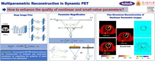
背景/目的正电子发射断层扫描(PET)的多参数成像由于动态数据的噪声和动力学参数的复杂映射而具有挑战性。虽然已经提出了直接参数重建等方法来改善图像质量,但局限性仍然存在,特别是对于非线性和小值微参数(例如,k2, k3)。本研究提出了一种新的无监督深度学习方法来重建和提高这些微参数的质量。方法将深度图像先验(DIP)与参数放大(PM)策略相结合,提出了一种直接参数化图像重建模型DIP-PM。该模型使用U-Net生成器预先使用CT图像预测多个参数图像,随后将每个输出通道放大一个因子以调整强度。利用测量投影数据与正演投影数据之间的对数似然损失对模型进行了优化。模拟两个示踪剂数据集进行评估:82Rb数据使用1组织室(1 TC)模型,18F-FDG数据使用2组织室(2 TC)模型,分别对1 TC k2和2 TC k3进行10倍放大。将DIP- pm与间接法、直接法(OTEM)和无参数放大的DIP法(DIP-only)进行比较。使用峰值信噪比(PSNR)、标准化均方根误差(NRMSE)和结构相似指数(SSIM)对模拟数据以及男性受试者的真实18F-FDG扫描进行性能评估。结果对于1 TC模型,OTEM法在K1重建中表现良好,但间接法和OTEM法在k2重建中均表现出高噪声和较差的效果。仅dip法抑制了k2的噪声,但未能重建心肌的精细结构。DIP-PM在细节结构保存良好的情况下优于其他方法,特别是在k2中,获得了最好的指标(PSNR: 19.00, NRMSE: 0.3002, SSIM: 0.9289)。对于2 TC模型,传统方法在估计所有非线性参数(K1, k2, k3)时存在高噪声和模糊结构,而基于dip的方法显著提高了图像质量。DIP-PM在k3上优于所有方法(PSNR: 21.89, NRMSE: 0.4054, SSIM: 0.8797),从而产生最准确的2 TC Ki图像(PSNR: 22.74, NRMSE: 0.4897, SSIM: 0.8391)。在实际FDG数据上,DIP-PM在估计K1、k2和k3方面也显示出明显的优势,同时保留了心肌结构。结论基于dip的直接参数成像在PET参数图像生成和质量改善方面具有一定的应用价值。研究表明,采用参数放大策略的DIP-PM方法可以提高非线性微参数图像的保真度。
本文章由计算机程序翻译,如有差异,请以英文原文为准。

Direct parametric reconstruction in dynamic PET using deep image prior and a novel parameter magnification strategy

Direct parametric reconstruction in dynamic PET using deep image prior and a novel parameter magnification strategy

Background/Purpose

Multiple parametric imaging in positron emission tomography (PET) is challenging due to the noisy dynamic data and the complex mapping to kinetic parameters. Although methods like direct parametric reconstruction have been proposed to improve the image quality, limitations persist, particularly for nonlinear and small-value micro-parameters (e.g., k2, k3). This study presents a novel unsupervised deep learning approach to reconstruct and improve the quality of these micro-parameters.

Methods

We proposed a direct parametric image reconstruction model, DIP-PM, integrating deep image prior (DIP) with a parameter magnification (PM) strategy. The model employs a U-Net generator to predict multiple parametric images using a CT image prior, with each output channel subsequently magnified by a factor to adjust the intensity. The model was optimized with a log-likelihood loss computed between the measured projection data and forward projected data. Two tracer datasets were simulated for evaluation: 82Rb data using the 1-tissue compartment (1 TC) model and 18F-FDG data using the 2-tissue compartment (2 TC) model, with 10-fold magnification applied to the 1 TC k2 and the 2 TC k3, respectively. DIP-PM was compared to the indirect method, direct algorithm (OTEM) and the DIP method without parameter magnification (DIP-only). Performance was assessed on phantom data using peak signal-to-noise ratio (PSNR), normalized root mean square error (NRMSE) and structural similarity index (SSIM), as well as on real 18F-FDG scan from a male subject.

Results

For the 1 TC model, OTEM performed well in K1 reconstruction, but both indirect and OTEM methods showed high noise and poor performance in k2. The DIP-only method suppressed noise in k2, but failed to reconstruct fine structures in the myocardium. DIP-PM outperformed other methods with well-preserved detailed structures, particularly in k2, achieving the best metrics (PSNR: 19.00, NRMSE: 0.3002, SSIM: 0.9289). For the 2 TC model, traditional methods exhibited high noise and blurred structures in estimating all nonlinear parameters (K1, k2, k3), while DIP-based methods significantly improved image quality. DIP-PM outperformed all methods in k3 (PSNR: 21.89, NRMSE: 0.4054, SSIM: 0.8797), and consequently produced the most accurate 2 TC Ki images (PSNR: 22.74, NRMSE: 0.4897, SSIM: 0.8391). On real FDG data, DIP-PM also showed evident advantages in estimating K1, k2 and k3 while preserving myocardial structures.

Conclusions

The results underscore the efficacy of the DIP-based direct parametric imaging in generating and improving quality of PET parametric images. This study suggests that the proposed DIP-PM method with the parameter magnification strategy can enhance the fidelity of nonlinear micro-parameter images.
求助全文
通过发布文献求助,成功后即可免费获取论文全文。 去求助
来源期刊
Computers in biology and medicine
Computers in biology and medicine 工程技术-工程:生物医学
CiteScore
11.70
自引率
10.40%
发文量
1086
审稿时长
74 days
期刊介绍: Computers in Biology and Medicine is an international forum for sharing groundbreaking advancements in the use of computers in bioscience and medicine. This journal serves as a medium for communicating essential research, instruction, ideas, and information regarding the rapidly evolving field of computer applications in these domains. By encouraging the exchange of knowledge, we aim to facilitate progress and innovation in the utilization of computers in biology and medicine.
×
引用
GB/T 7714-2015
复制
MLA
复制
APA
复制
导出至
BibTeX EndNote RefMan NoteFirst NoteExpress
×
提示
您的信息不完整,为了账户安全,请先补充。
现在去补充
×
提示
您因"违规操作"
具体请查看互助需知
我知道了
×
提示
确定
请完成安全验证×
copy
已复制链接
快去分享给好友吧!
我知道了
右上角分享
点击右上角分享
0
联系我们:info@booksci.cn Book学术提供免费学术资源搜索服务,方便国内外学者检索中英文文献。致力于提供最便捷和优质的服务体验。 Copyright © 2023 布克学术 All rights reserved.
京ICP备2023020795号-1
ghs 京公网安备 11010802042870号
Book学术文献互助
Book学术文献互助群
群 号:604180095
Book学术官方微信