Ewelina Czuba-Pakuła, Jolanta Ochocińska, Sebastian Głowiński, Alicja Braczko, Ryszard T Smoleński, Grażyna Lietzau, Przemysław Kowiański
{"title":"高胆固醇血症持续时间和脑面积决定Apo E-/-/LDLR-/-双敲除小鼠的炎症反应强度和凋亡介质激活","authors":"Ewelina Czuba-Pakuła, Jolanta Ochocińska, Sebastian Głowiński, Alicja Braczko, Ryszard T Smoleński, Grażyna Lietzau, Przemysław Kowiański","doi":"10.1007/s10571-025-01562-0","DOIUrl":null,"url":null,"abstract":"<p><p>Hypercholesterolemia (Hch) is a risk factor for cerebrovascular and neurodegenerative diseases, manifesting with symptoms that vary depending on damage to specific brain regions. Hch triggers inflammatory responses and cell death. However, the progression of these processes in relation to the duration of Hch and the location of pathology in the central nervous system remains unclear. Therefore, we aimed to investigate (1) the impact of age and duration of Hch on neuroinflammatory responses and programmed cell death in the brain and (2) the intensity of these processes in various brain areas during Hch. In this study, we used 3-, 6-, and 12-month-old male Apo E<sup>-/-</sup>/LDLR<sup>-/-</sup> double-knockout mice and age-matched wild-type C57BL/6 mice (control group). Concentrations of cytokines IL-1β, IL-4, and IL-6, as well as apoptotic mediators AIF and Cas-3, were measured using enzyme-linked immunosorbent assay in the whole brain and separately in the prefrontal cortex (PFCx), hippocampus (HIP), and striatum (STR). The results showed that the Hch-induced release of cytokines IL-1β and IL-6, decreased expression of IL-4, and elevated level of apoptotic markers AIF and Cas-3 correlated with Hch duration. The inflammatory response and expression of apoptotic markers were more pronounced in the HIP and STR compared to the PFCx. Our results indicate a correlation between the neurodegenerative effects of Hch and its duration and highlight the varying susceptibility of different brain areas to Hch-induced damage.</p>","PeriodicalId":9742,"journal":{"name":"Cellular and Molecular Neurobiology","volume":"45 1","pages":"55"},"PeriodicalIF":4.8000,"publicationDate":"2025-05-30","publicationTypes":"Journal Article","fieldsOfStudy":null,"isOpenAccess":false,"openAccessPdf":"https://www.ncbi.nlm.nih.gov/pmc/articles/PMC12125462/pdf/","citationCount":"0","resultStr":"{\"title\":\"Hypercholesterolemia Duration and Brain Area Determine Inflammatory Response Intensity and Apoptotic Mediator Activation in Apo E<sup>-/-</sup>/LDLR<sup>-/-</sup> Double-Knockout Mice.\",\"authors\":\"Ewelina Czuba-Pakuła, Jolanta Ochocińska, Sebastian Głowiński, Alicja Braczko, Ryszard T Smoleński, Grażyna Lietzau, Przemysław Kowiański\",\"doi\":\"10.1007/s10571-025-01562-0\",\"DOIUrl\":null,\"url\":null,\"abstract\":\"<p><p>Hypercholesterolemia (Hch) is a risk factor for cerebrovascular and neurodegenerative diseases, manifesting with symptoms that vary depending on damage to specific brain regions. Hch triggers inflammatory responses and cell death. However, the progression of these processes in relation to the duration of Hch and the location of pathology in the central nervous system remains unclear. Therefore, we aimed to investigate (1) the impact of age and duration of Hch on neuroinflammatory responses and programmed cell death in the brain and (2) the intensity of these processes in various brain areas during Hch. In this study, we used 3-, 6-, and 12-month-old male Apo E<sup>-/-</sup>/LDLR<sup>-/-</sup> double-knockout mice and age-matched wild-type C57BL/6 mice (control group). Concentrations of cytokines IL-1β, IL-4, and IL-6, as well as apoptotic mediators AIF and Cas-3, were measured using enzyme-linked immunosorbent assay in the whole brain and separately in the prefrontal cortex (PFCx), hippocampus (HIP), and striatum (STR). The results showed that the Hch-induced release of cytokines IL-1β and IL-6, decreased expression of IL-4, and elevated level of apoptotic markers AIF and Cas-3 correlated with Hch duration. The inflammatory response and expression of apoptotic markers were more pronounced in the HIP and STR compared to the PFCx. Our results indicate a correlation between the neurodegenerative effects of Hch and its duration and highlight the varying susceptibility of different brain areas to Hch-induced damage.</p>\",\"PeriodicalId\":9742,\"journal\":{\"name\":\"Cellular and Molecular Neurobiology\",\"volume\":\"45 1\",\"pages\":\"55\"},\"PeriodicalIF\":4.8000,\"publicationDate\":\"2025-05-30\",\"publicationTypes\":\"Journal Article\",\"fieldsOfStudy\":null,\"isOpenAccess\":false,\"openAccessPdf\":\"https://www.ncbi.nlm.nih.gov/pmc/articles/PMC12125462/pdf/\",\"citationCount\":\"0\",\"resultStr\":null,\"platform\":\"Semanticscholar\",\"paperid\":null,\"PeriodicalName\":\"Cellular and Molecular Neurobiology\",\"FirstCategoryId\":\"3\",\"ListUrlMain\":\"https://doi.org/10.1007/s10571-025-01562-0\",\"RegionNum\":4,\"RegionCategory\":\"医学\",\"ArticlePicture\":[],\"TitleCN\":null,\"AbstractTextCN\":null,\"PMCID\":null,\"EPubDate\":\"\",\"PubModel\":\"\",\"JCR\":\"Q3\",\"JCRName\":\"CELL BIOLOGY\",\"Score\":null,\"Total\":0}","platform":"Semanticscholar","paperid":null,"PeriodicalName":"Cellular and Molecular Neurobiology","FirstCategoryId":"3","ListUrlMain":"https://doi.org/10.1007/s10571-025-01562-0","RegionNum":4,"RegionCategory":"医学","ArticlePicture":[],"TitleCN":null,"AbstractTextCN":null,"PMCID":null,"EPubDate":"","PubModel":"","JCR":"Q3","JCRName":"CELL BIOLOGY","Score":null,"Total":0}
Hypercholesterolemia Duration and Brain Area Determine Inflammatory Response Intensity and Apoptotic Mediator Activation in Apo E-/-/LDLR-/- Double-Knockout Mice.
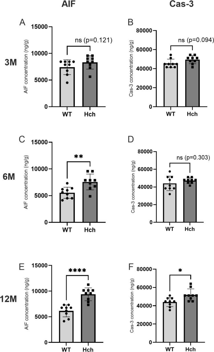
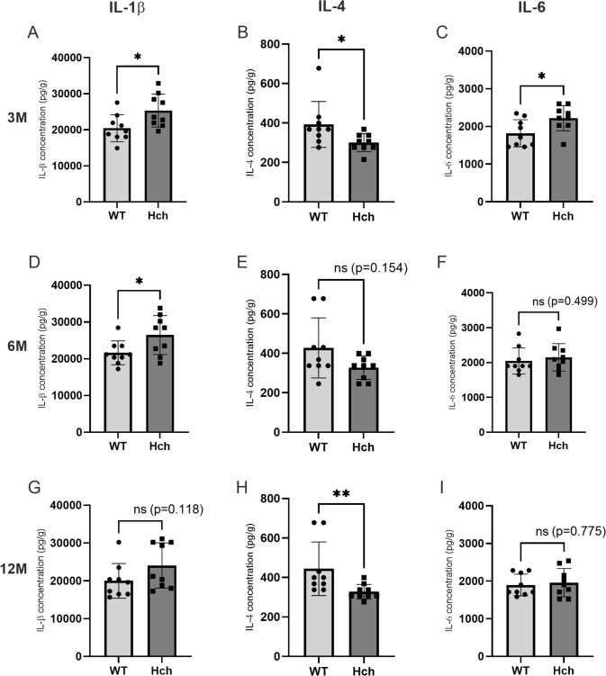
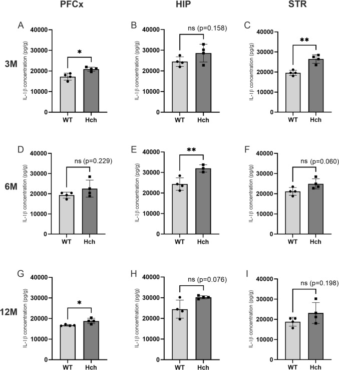
Hypercholesterolemia (Hch) is a risk factor for cerebrovascular and neurodegenerative diseases, manifesting with symptoms that vary depending on damage to specific brain regions. Hch triggers inflammatory responses and cell death. However, the progression of these processes in relation to the duration of Hch and the location of pathology in the central nervous system remains unclear. Therefore, we aimed to investigate (1) the impact of age and duration of Hch on neuroinflammatory responses and programmed cell death in the brain and (2) the intensity of these processes in various brain areas during Hch. In this study, we used 3-, 6-, and 12-month-old male Apo E-/-/LDLR-/- double-knockout mice and age-matched wild-type C57BL/6 mice (control group). Concentrations of cytokines IL-1β, IL-4, and IL-6, as well as apoptotic mediators AIF and Cas-3, were measured using enzyme-linked immunosorbent assay in the whole brain and separately in the prefrontal cortex (PFCx), hippocampus (HIP), and striatum (STR). The results showed that the Hch-induced release of cytokines IL-1β and IL-6, decreased expression of IL-4, and elevated level of apoptotic markers AIF and Cas-3 correlated with Hch duration. The inflammatory response and expression of apoptotic markers were more pronounced in the HIP and STR compared to the PFCx. Our results indicate a correlation between the neurodegenerative effects of Hch and its duration and highlight the varying susceptibility of different brain areas to Hch-induced damage.
期刊介绍:
Cellular and Molecular Neurobiology publishes original research concerned with the analysis of neuronal and brain function at the cellular and subcellular levels. The journal offers timely, peer-reviewed articles that describe anatomic, genetic, physiologic, pharmacologic, and biochemical approaches to the study of neuronal function and the analysis of elementary mechanisms. Studies are presented on isolated mammalian tissues and intact animals, with investigations aimed at the molecular mechanisms or neuronal responses at the level of single cells. Cellular and Molecular Neurobiology also presents studies of the effects of neurons on other organ systems, such as analysis of the electrical or biochemical response to neurotransmitters or neurohormones on smooth muscle or gland cells.
 求助内容:
求助内容: 应助结果提醒方式:


