Jun Liu, Huijuan Pan, Yong Bao, Li Huang, Yunyun Hu
{"title":"肌肉骨骼超声在中风后偏瘫肩关节康复中的临床应用。","authors":"Jun Liu, Huijuan Pan, Yong Bao, Li Huang, Yunyun Hu","doi":"10.3389/fresc.2025.1576890","DOIUrl":null,"url":null,"abstract":"<p><strong>Objective: </strong>This study aimed to assess the utility of musculoskeletal ultrasound (MSUS) in the rehabilitation of stroke patients with hemiplegic shoulder pain.</p><p><strong>Methods: </strong>We conducted a study involving 80 stroke patients with hemiplegia and concomitant shoulder pain on the affected side, admitted to our hospital between April 2020 and March 2021. MSUS was used to evaluate shoulder structures, including the long head of the biceps brachii tendon (BICT) and its sheath, rotator cuff, subacromial-subdeltoid (SA-SD) bursa, labrum, acromioclavicular ligament, acromiocoracoid ligament, and acromion-greater tuberosity (AGT) distance. We compared pre- and post-rehabilitation measurements of supraspinatus tendon (SST) thickness, BICT sheath effusion thickness, SA-SD bursa effusion thickness, AGT distance, and visual analog scale (VAS) scores. Statistical significance was set at <i>P</i> < 0.05.</p><p><strong>Results: </strong>Post-rehabilitation, the SST thickness on the hemiplegic side showed a statistically significant reduction (<i>P</i> = 0.023). No significant difference was observed in the mean maximum rupture diameter (<i>P</i> = 0.796). Both BICT sheath effusion (<i>P</i> < 0.001) and SA-SD bursa effusion (<i>P</i> < 0.001) exhibited significant decreases. The AGT distance on the hemiplegic side also demonstrated a statistically significant change (<i>P</i> < 0.001). Additionally, the VAS score significantly improved post-rehabilitation (<i>P</i> < 0.001).</p><p><strong>Conclusion: </strong>MSUS is a feasible and reproducible tool for monitoring rehabilitation progress in stroke patients with hemiplegic shoulder pain.</p>","PeriodicalId":73102,"journal":{"name":"Frontiers in rehabilitation sciences","volume":"6 ","pages":"1576890"},"PeriodicalIF":1.9000,"publicationDate":"2025-05-15","publicationTypes":"Journal Article","fieldsOfStudy":null,"isOpenAccess":false,"openAccessPdf":"https://www.ncbi.nlm.nih.gov/pmc/articles/PMC12119546/pdf/","citationCount":"0","resultStr":"{\"title\":\"The clinical utility of musculoskeletal ultrasonography in hemiplegic shoulder rehabilitation poststroke.\",\"authors\":\"Jun Liu, Huijuan Pan, Yong Bao, Li Huang, Yunyun Hu\",\"doi\":\"10.3389/fresc.2025.1576890\",\"DOIUrl\":null,\"url\":null,\"abstract\":\"<p><strong>Objective: </strong>This study aimed to assess the utility of musculoskeletal ultrasound (MSUS) in the rehabilitation of stroke patients with hemiplegic shoulder pain.</p><p><strong>Methods: </strong>We conducted a study involving 80 stroke patients with hemiplegia and concomitant shoulder pain on the affected side, admitted to our hospital between April 2020 and March 2021. MSUS was used to evaluate shoulder structures, including the long head of the biceps brachii tendon (BICT) and its sheath, rotator cuff, subacromial-subdeltoid (SA-SD) bursa, labrum, acromioclavicular ligament, acromiocoracoid ligament, and acromion-greater tuberosity (AGT) distance. We compared pre- and post-rehabilitation measurements of supraspinatus tendon (SST) thickness, BICT sheath effusion thickness, SA-SD bursa effusion thickness, AGT distance, and visual analog scale (VAS) scores. Statistical significance was set at <i>P</i> < 0.05.</p><p><strong>Results: </strong>Post-rehabilitation, the SST thickness on the hemiplegic side showed a statistically significant reduction (<i>P</i> = 0.023). No significant difference was observed in the mean maximum rupture diameter (<i>P</i> = 0.796). Both BICT sheath effusion (<i>P</i> < 0.001) and SA-SD bursa effusion (<i>P</i> < 0.001) exhibited significant decreases. The AGT distance on the hemiplegic side also demonstrated a statistically significant change (<i>P</i> < 0.001). Additionally, the VAS score significantly improved post-rehabilitation (<i>P</i> < 0.001).</p><p><strong>Conclusion: </strong>MSUS is a feasible and reproducible tool for monitoring rehabilitation progress in stroke patients with hemiplegic shoulder pain.</p>\",\"PeriodicalId\":73102,\"journal\":{\"name\":\"Frontiers in rehabilitation sciences\",\"volume\":\"6 \",\"pages\":\"1576890\"},\"PeriodicalIF\":1.9000,\"publicationDate\":\"2025-05-15\",\"publicationTypes\":\"Journal Article\",\"fieldsOfStudy\":null,\"isOpenAccess\":false,\"openAccessPdf\":\"https://www.ncbi.nlm.nih.gov/pmc/articles/PMC12119546/pdf/\",\"citationCount\":\"0\",\"resultStr\":null,\"platform\":\"Semanticscholar\",\"paperid\":null,\"PeriodicalName\":\"Frontiers in rehabilitation sciences\",\"FirstCategoryId\":\"1085\",\"ListUrlMain\":\"https://doi.org/10.3389/fresc.2025.1576890\",\"RegionNum\":0,\"RegionCategory\":null,\"ArticlePicture\":[],\"TitleCN\":null,\"AbstractTextCN\":null,\"PMCID\":null,\"EPubDate\":\"2025/1/1 0:00:00\",\"PubModel\":\"eCollection\",\"JCR\":\"Q3\",\"JCRName\":\"REHABILITATION\",\"Score\":null,\"Total\":0}","platform":"Semanticscholar","paperid":null,"PeriodicalName":"Frontiers in rehabilitation sciences","FirstCategoryId":"1085","ListUrlMain":"https://doi.org/10.3389/fresc.2025.1576890","RegionNum":0,"RegionCategory":null,"ArticlePicture":[],"TitleCN":null,"AbstractTextCN":null,"PMCID":null,"EPubDate":"2025/1/1 0:00:00","PubModel":"eCollection","JCR":"Q3","JCRName":"REHABILITATION","Score":null,"Total":0}
The clinical utility of musculoskeletal ultrasonography in hemiplegic shoulder rehabilitation poststroke.
Objective: This study aimed to assess the utility of musculoskeletal ultrasound (MSUS) in the rehabilitation of stroke patients with hemiplegic shoulder pain.
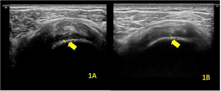
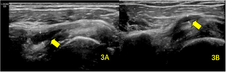
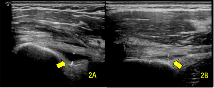
Methods: We conducted a study involving 80 stroke patients with hemiplegia and concomitant shoulder pain on the affected side, admitted to our hospital between April 2020 and March 2021. MSUS was used to evaluate shoulder structures, including the long head of the biceps brachii tendon (BICT) and its sheath, rotator cuff, subacromial-subdeltoid (SA-SD) bursa, labrum, acromioclavicular ligament, acromiocoracoid ligament, and acromion-greater tuberosity (AGT) distance. We compared pre- and post-rehabilitation measurements of supraspinatus tendon (SST) thickness, BICT sheath effusion thickness, SA-SD bursa effusion thickness, AGT distance, and visual analog scale (VAS) scores. Statistical significance was set at P < 0.05.
Results: Post-rehabilitation, the SST thickness on the hemiplegic side showed a statistically significant reduction (P = 0.023). No significant difference was observed in the mean maximum rupture diameter (P = 0.796). Both BICT sheath effusion (P < 0.001) and SA-SD bursa effusion (P < 0.001) exhibited significant decreases. The AGT distance on the hemiplegic side also demonstrated a statistically significant change (P < 0.001). Additionally, the VAS score significantly improved post-rehabilitation (P < 0.001).
Conclusion: MSUS is a feasible and reproducible tool for monitoring rehabilitation progress in stroke patients with hemiplegic shoulder pain.

 求助内容:
求助内容: 应助结果提醒方式:


