Reham Gonnah, Julia E. Parker, Robert P. W. Davies and Maisoon Al-Jawad
{"title":"同步x射线纳米探针和相关电子显微镜揭示了自组装肽在磷酸钙成核中的表面化学作用。","authors":"Reham Gonnah, Julia E. Parker, Robert P. W. Davies and Maisoon Al-Jawad","doi":"10.1039/D5FD00017C","DOIUrl":null,"url":null,"abstract":"<p >A biomimetic peptide (P<small><sub>11</sub></small>-4), which is predominantly negatively-charged, facilitates the nucleation of hydroxyapatite (HAp). P<small><sub>11</sub></small>-4 self-assembles into fibrils <em>via</em> β-sheet formation, creating a 3D-gel-network. Here, X-ray nanoimaging and correlative scanning electron microscopy (SEM) investigated P<small><sub>11</sub></small>-4's surface chemistry and its ability to nucleate HAp in the absence of the 3D-gel-network. P<small><sub>11</sub></small>-4 was deposited on silicon nitride (SiN) windows, which were immersed in a mineralising solution (MS) and then mapped using nano-X-ray fluorescence (n-XRF) and differential phase contrast imaging at the hard X-ray nanoprobe beamline (I14) at Diamond Light Source. Elemental calcium and phosphorus maps were extracted using n-XRF, and compared with and without P<small><sub>11</sub></small>-4. The windows were subsequently mapped using SEM and Energy Dispersive Spectroscopy (EDS) to confirm the morphology and elemental compositions of the formed structures. The calcium : phosphorus ratios were calculated to identify the phases formed. P<small><sub>11</sub></small>-4 increased the calcium and phosphorus signals with time in MS compared to the control (without P<small><sub>11</sub></small>-4). After 12 hours in MS, calcium ions accumulated on the deposited β-sheets, attracting phosphorus ions at later time points. From the morphology in the images and EDS analysis, the spherical calcium phosphate (CaP) structures appeared to be amorphous, indicating the formation of precursors, likely amorphous CaP, at early time points. In the presence of P<small><sub>11</sub></small>-4, these structures grew and fused into larger CaP formations over time, unlike in the control. Nano-imaging techniques highlighted that P<small><sub>11</sub></small>-4's surface chemistry accelerates the kinetics and controls the initial CaP crystallisation process, resulting in an amorphous CaP phase.</p>","PeriodicalId":49075,"journal":{"name":"Faraday Discussions","volume":"261 ","pages":" 132-150"},"PeriodicalIF":3.1000,"publicationDate":"2025-03-19","publicationTypes":"Journal Article","fieldsOfStudy":null,"isOpenAccess":false,"openAccessPdf":"https://pubs.rsc.org/en/content/articlepdf/2025/fd/d5fd00017c?page=search","citationCount":"0","resultStr":"{\"title\":\"Synchrotron X-ray nanoprobe and correlative electron microscopy reveal the role of surface chemistry of self-assembling peptides in calcium phosphate nucleation†\",\"authors\":\"Reham Gonnah, Julia E. Parker, Robert P. W. Davies and Maisoon Al-Jawad\",\"doi\":\"10.1039/D5FD00017C\",\"DOIUrl\":null,\"url\":null,\"abstract\":\"<p >A biomimetic peptide (P<small><sub>11</sub></small>-4), which is predominantly negatively-charged, facilitates the nucleation of hydroxyapatite (HAp). P<small><sub>11</sub></small>-4 self-assembles into fibrils <em>via</em> β-sheet formation, creating a 3D-gel-network. Here, X-ray nanoimaging and correlative scanning electron microscopy (SEM) investigated P<small><sub>11</sub></small>-4's surface chemistry and its ability to nucleate HAp in the absence of the 3D-gel-network. P<small><sub>11</sub></small>-4 was deposited on silicon nitride (SiN) windows, which were immersed in a mineralising solution (MS) and then mapped using nano-X-ray fluorescence (n-XRF) and differential phase contrast imaging at the hard X-ray nanoprobe beamline (I14) at Diamond Light Source. Elemental calcium and phosphorus maps were extracted using n-XRF, and compared with and without P<small><sub>11</sub></small>-4. The windows were subsequently mapped using SEM and Energy Dispersive Spectroscopy (EDS) to confirm the morphology and elemental compositions of the formed structures. The calcium : phosphorus ratios were calculated to identify the phases formed. P<small><sub>11</sub></small>-4 increased the calcium and phosphorus signals with time in MS compared to the control (without P<small><sub>11</sub></small>-4). After 12 hours in MS, calcium ions accumulated on the deposited β-sheets, attracting phosphorus ions at later time points. From the morphology in the images and EDS analysis, the spherical calcium phosphate (CaP) structures appeared to be amorphous, indicating the formation of precursors, likely amorphous CaP, at early time points. In the presence of P<small><sub>11</sub></small>-4, these structures grew and fused into larger CaP formations over time, unlike in the control. Nano-imaging techniques highlighted that P<small><sub>11</sub></small>-4's surface chemistry accelerates the kinetics and controls the initial CaP crystallisation process, resulting in an amorphous CaP phase.</p>\",\"PeriodicalId\":49075,\"journal\":{\"name\":\"Faraday Discussions\",\"volume\":\"261 \",\"pages\":\" 132-150\"},\"PeriodicalIF\":3.1000,\"publicationDate\":\"2025-03-19\",\"publicationTypes\":\"Journal Article\",\"fieldsOfStudy\":null,\"isOpenAccess\":false,\"openAccessPdf\":\"https://pubs.rsc.org/en/content/articlepdf/2025/fd/d5fd00017c?page=search\",\"citationCount\":\"0\",\"resultStr\":null,\"platform\":\"Semanticscholar\",\"paperid\":null,\"PeriodicalName\":\"Faraday Discussions\",\"FirstCategoryId\":\"92\",\"ListUrlMain\":\"https://pubs.rsc.org/en/content/articlelanding/2025/fd/d5fd00017c\",\"RegionNum\":3,\"RegionCategory\":\"化学\",\"ArticlePicture\":[],\"TitleCN\":null,\"AbstractTextCN\":null,\"PMCID\":null,\"EPubDate\":\"\",\"PubModel\":\"\",\"JCR\":\"Q2\",\"JCRName\":\"Chemistry\",\"Score\":null,\"Total\":0}","platform":"Semanticscholar","paperid":null,"PeriodicalName":"Faraday Discussions","FirstCategoryId":"92","ListUrlMain":"https://pubs.rsc.org/en/content/articlelanding/2025/fd/d5fd00017c","RegionNum":3,"RegionCategory":"化学","ArticlePicture":[],"TitleCN":null,"AbstractTextCN":null,"PMCID":null,"EPubDate":"","PubModel":"","JCR":"Q2","JCRName":"Chemistry","Score":null,"Total":0}
Synchrotron X-ray nanoprobe and correlative electron microscopy reveal the role of surface chemistry of self-assembling peptides in calcium phosphate nucleation†
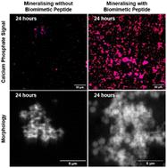
A biomimetic peptide (P11-4), which is predominantly negatively-charged, facilitates the nucleation of hydroxyapatite (HAp). P11-4 self-assembles into fibrils via β-sheet formation, creating a 3D-gel-network. Here, X-ray nanoimaging and correlative scanning electron microscopy (SEM) investigated P11-4's surface chemistry and its ability to nucleate HAp in the absence of the 3D-gel-network. P11-4 was deposited on silicon nitride (SiN) windows, which were immersed in a mineralising solution (MS) and then mapped using nano-X-ray fluorescence (n-XRF) and differential phase contrast imaging at the hard X-ray nanoprobe beamline (I14) at Diamond Light Source. Elemental calcium and phosphorus maps were extracted using n-XRF, and compared with and without P11-4. The windows were subsequently mapped using SEM and Energy Dispersive Spectroscopy (EDS) to confirm the morphology and elemental compositions of the formed structures. The calcium : phosphorus ratios were calculated to identify the phases formed. P11-4 increased the calcium and phosphorus signals with time in MS compared to the control (without P11-4). After 12 hours in MS, calcium ions accumulated on the deposited β-sheets, attracting phosphorus ions at later time points. From the morphology in the images and EDS analysis, the spherical calcium phosphate (CaP) structures appeared to be amorphous, indicating the formation of precursors, likely amorphous CaP, at early time points. In the presence of P11-4, these structures grew and fused into larger CaP formations over time, unlike in the control. Nano-imaging techniques highlighted that P11-4's surface chemistry accelerates the kinetics and controls the initial CaP crystallisation process, resulting in an amorphous CaP phase.
 求助内容:
求助内容: 应助结果提醒方式:


