Lise Guichaoua, Natalie Reznikov, Bryce D. Stewart, Roland Kröger and Raynald Gauvin
{"title":"王扇贝(Pecten maximus)壳的SEM, EBSD和拉曼光谱联合晶体学研究。","authors":"Lise Guichaoua, Natalie Reznikov, Bryce D. Stewart, Roland Kröger and Raynald Gauvin","doi":"10.1039/D5FD00029G","DOIUrl":null,"url":null,"abstract":"<p >The shells of <em>Pecten maximus</em> (king scallop) are composed primarily of polycrystalline calcitic calcium carbonate, with a crucial aragonitic prismatic myostracum layer that facilitates soft tissue attachment and contributes to mechanical strength. Despite its importance, the impact of environmental stressors, such as metal contamination, on the myostracum remains underexplored. Hence, this study's main goal was to shed light on the microstructure and crystallography of king scallop shells, particularly the myostracum region, using a combination of scanning electron microscopy (SEM), electron backscatter diffraction (EBSD) analysis, and Raman spectroscopy. This approach was chosen to develop new protocols that integrate imaging techniques for a systematic analysis of pollution effects on shell growth and properties. Such an understanding is crucial for assessing the impact of environmental contamination on shell structure and composition. We compared samples from a metal contaminated site (Laxey) with those from non-contaminated site (Bradda) around the Isle of Man, to determine which structural and crystallographic information is detectable using the selected microscopy and spectroscopy techniques. SEM imaging showed a similar myostracum organization in specimens from both sites, with elongated, oriented grains. However, the non-contaminated site shell had more regular and elongated grains, while the shell from the contaminated site exhibited a broader grain size distribution, visible <em>via</em> electron channeling contrast and backscattered electron detection. EBSD analysis confirmed that both types exhibited well-crystallized aragonite in the myostracum, with slight differences in grain orientation and grain size with co-orientation indicating a marginal reduction in crystallographic alignment in the contaminated site shell. Raman maps reveal shifts in peak positions, indicating crystallite strain and differences in grain size. These variations may be related to a biological adaptation aimed at toughening the shell, but pollution could disrupt this crystallization process, weakening the shell. The combination of these techniques can advance our understanding of the microstructural alterations caused by metal contamination, highlighting its potential impact on the structural integrity of the shell. This study is a proof of principle study showing how a combination of different established imaging techniques can provide complementary and novel insights into the structure and composition of the king scallop myostracum. This systematic approach aims to develop new evaluation approaches for the study of the effects of environmental pollutants on the crystallography and microstructure of marine bivalve shells and hence their resilience.</p>","PeriodicalId":49075,"journal":{"name":"Faraday Discussions","volume":"261 ","pages":" 484-500"},"PeriodicalIF":3.1000,"publicationDate":"2025-02-28","publicationTypes":"Journal Article","fieldsOfStudy":null,"isOpenAccess":false,"openAccessPdf":"https://pubs.rsc.org/en/content/articlepdf/2025/fd/d5fd00029g?page=search","citationCount":"0","resultStr":"{\"title\":\"Combined crystallographic study of king scallop (Pecten maximus) shells using SEM, EBSD and Raman spectroscopy\",\"authors\":\"Lise Guichaoua, Natalie Reznikov, Bryce D. Stewart, Roland Kröger and Raynald Gauvin\",\"doi\":\"10.1039/D5FD00029G\",\"DOIUrl\":null,\"url\":null,\"abstract\":\"<p >The shells of <em>Pecten maximus</em> (king scallop) are composed primarily of polycrystalline calcitic calcium carbonate, with a crucial aragonitic prismatic myostracum layer that facilitates soft tissue attachment and contributes to mechanical strength. Despite its importance, the impact of environmental stressors, such as metal contamination, on the myostracum remains underexplored. Hence, this study's main goal was to shed light on the microstructure and crystallography of king scallop shells, particularly the myostracum region, using a combination of scanning electron microscopy (SEM), electron backscatter diffraction (EBSD) analysis, and Raman spectroscopy. This approach was chosen to develop new protocols that integrate imaging techniques for a systematic analysis of pollution effects on shell growth and properties. Such an understanding is crucial for assessing the impact of environmental contamination on shell structure and composition. We compared samples from a metal contaminated site (Laxey) with those from non-contaminated site (Bradda) around the Isle of Man, to determine which structural and crystallographic information is detectable using the selected microscopy and spectroscopy techniques. SEM imaging showed a similar myostracum organization in specimens from both sites, with elongated, oriented grains. However, the non-contaminated site shell had more regular and elongated grains, while the shell from the contaminated site exhibited a broader grain size distribution, visible <em>via</em> electron channeling contrast and backscattered electron detection. EBSD analysis confirmed that both types exhibited well-crystallized aragonite in the myostracum, with slight differences in grain orientation and grain size with co-orientation indicating a marginal reduction in crystallographic alignment in the contaminated site shell. Raman maps reveal shifts in peak positions, indicating crystallite strain and differences in grain size. These variations may be related to a biological adaptation aimed at toughening the shell, but pollution could disrupt this crystallization process, weakening the shell. The combination of these techniques can advance our understanding of the microstructural alterations caused by metal contamination, highlighting its potential impact on the structural integrity of the shell. This study is a proof of principle study showing how a combination of different established imaging techniques can provide complementary and novel insights into the structure and composition of the king scallop myostracum. This systematic approach aims to develop new evaluation approaches for the study of the effects of environmental pollutants on the crystallography and microstructure of marine bivalve shells and hence their resilience.</p>\",\"PeriodicalId\":49075,\"journal\":{\"name\":\"Faraday Discussions\",\"volume\":\"261 \",\"pages\":\" 484-500\"},\"PeriodicalIF\":3.1000,\"publicationDate\":\"2025-02-28\",\"publicationTypes\":\"Journal Article\",\"fieldsOfStudy\":null,\"isOpenAccess\":false,\"openAccessPdf\":\"https://pubs.rsc.org/en/content/articlepdf/2025/fd/d5fd00029g?page=search\",\"citationCount\":\"0\",\"resultStr\":null,\"platform\":\"Semanticscholar\",\"paperid\":null,\"PeriodicalName\":\"Faraday Discussions\",\"FirstCategoryId\":\"92\",\"ListUrlMain\":\"https://pubs.rsc.org/en/content/articlelanding/2025/fd/d5fd00029g\",\"RegionNum\":3,\"RegionCategory\":\"化学\",\"ArticlePicture\":[],\"TitleCN\":null,\"AbstractTextCN\":null,\"PMCID\":null,\"EPubDate\":\"\",\"PubModel\":\"\",\"JCR\":\"Q2\",\"JCRName\":\"Chemistry\",\"Score\":null,\"Total\":0}","platform":"Semanticscholar","paperid":null,"PeriodicalName":"Faraday Discussions","FirstCategoryId":"92","ListUrlMain":"https://pubs.rsc.org/en/content/articlelanding/2025/fd/d5fd00029g","RegionNum":3,"RegionCategory":"化学","ArticlePicture":[],"TitleCN":null,"AbstractTextCN":null,"PMCID":null,"EPubDate":"","PubModel":"","JCR":"Q2","JCRName":"Chemistry","Score":null,"Total":0}
Combined crystallographic study of king scallop (Pecten maximus) shells using SEM, EBSD and Raman spectroscopy
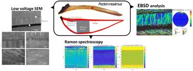
The shells of Pecten maximus (king scallop) are composed primarily of polycrystalline calcitic calcium carbonate, with a crucial aragonitic prismatic myostracum layer that facilitates soft tissue attachment and contributes to mechanical strength. Despite its importance, the impact of environmental stressors, such as metal contamination, on the myostracum remains underexplored. Hence, this study's main goal was to shed light on the microstructure and crystallography of king scallop shells, particularly the myostracum region, using a combination of scanning electron microscopy (SEM), electron backscatter diffraction (EBSD) analysis, and Raman spectroscopy. This approach was chosen to develop new protocols that integrate imaging techniques for a systematic analysis of pollution effects on shell growth and properties. Such an understanding is crucial for assessing the impact of environmental contamination on shell structure and composition. We compared samples from a metal contaminated site (Laxey) with those from non-contaminated site (Bradda) around the Isle of Man, to determine which structural and crystallographic information is detectable using the selected microscopy and spectroscopy techniques. SEM imaging showed a similar myostracum organization in specimens from both sites, with elongated, oriented grains. However, the non-contaminated site shell had more regular and elongated grains, while the shell from the contaminated site exhibited a broader grain size distribution, visible via electron channeling contrast and backscattered electron detection. EBSD analysis confirmed that both types exhibited well-crystallized aragonite in the myostracum, with slight differences in grain orientation and grain size with co-orientation indicating a marginal reduction in crystallographic alignment in the contaminated site shell. Raman maps reveal shifts in peak positions, indicating crystallite strain and differences in grain size. These variations may be related to a biological adaptation aimed at toughening the shell, but pollution could disrupt this crystallization process, weakening the shell. The combination of these techniques can advance our understanding of the microstructural alterations caused by metal contamination, highlighting its potential impact on the structural integrity of the shell. This study is a proof of principle study showing how a combination of different established imaging techniques can provide complementary and novel insights into the structure and composition of the king scallop myostracum. This systematic approach aims to develop new evaluation approaches for the study of the effects of environmental pollutants on the crystallography and microstructure of marine bivalve shells and hence their resilience.
 求助内容:
求助内容: 应助结果提醒方式:


