Seungwoo Cha, Kyoung Tae Kim, Won Kee Chang, Nam-Jong Paik, Ji Soo Choi, Hyunmi Lim, Won-Seok Kim, Jeonghun Ku
{"title":"基于脑电图的运动图像神经反馈对脑卒中患者运动尝试中mu抑制的影响。","authors":"Seungwoo Cha, Kyoung Tae Kim, Won Kee Chang, Nam-Jong Paik, Ji Soo Choi, Hyunmi Lim, Won-Seok Kim, Jeonghun Ku","doi":"10.1186/s12984-025-01653-5","DOIUrl":null,"url":null,"abstract":"<p><strong>Objective: </strong>The primary aim of this study was to explore the neurophysiological effects of motor imagery neurofeedback using electroencephalography (EEG), specifically focusing on mu suppression during serial motor attempts, and to assess its potential benefits in patients with subacute stroke.</p><p><strong>Methods: </strong>A total of 15 patients with hemiplegia following subacute ischemic stroke were prospectively enrolled in this randomized cross-over study. This study comprised two experiments: neurofeedback and sham. Each experiment included four blocks: three blocks of resting, grasp, resting, and an interventional task, followed by one block of resting and grasp. During the resting sessions, participants fixated on a white cross on a black background for 2 min without moving their upper extremities. In the grasp sessions, participants were instructed to grasp and release their paretic hand at a frequency of about 1 Hz for 3 min while maintaining fixation on the white cross. During the interventional task, the neurofeedback presented a punching image using the affected upper limb, corresponding to the mu suppression induced by imagined movement for 3 min. In contrast, the sham presented an image based on mu suppression data from randomly selected participants. EEG data were recorded throughout the experiment, and data from electrodes C3/C4 and P3/P4 were analyzed to compare the degree of mu suppression between the neurofeedback and sham experiments.</p><p><strong>Results: </strong>Significant mu suppression was observed in the bilateral motor and parietal cortices during the neurofeedback experiment compared with the sham across serial sessions (p < 0.001). Following neurofeedback, real grasping sessions showed progressive strengthening of mu suppression in the ipsilesional motor cortex and bilateral parietal cortices compared to sessions following sham (p < 0.05). This effect was not observed in the contralesional motor cortex.</p><p><strong>Conclusions: </strong>Motor imagery neurofeedback significantly enhances mu suppression in the ipsilesional motor and bilateral parietal cortices during motor attempts in patients with subacute stroke. These findings suggest that motor imagery neurofeedback could serve as a promising adjunctive therapy to enhance motor-related cortical activity and support motor rehabilitation in patients with stroke.</p>","PeriodicalId":16384,"journal":{"name":"Journal of NeuroEngineering and Rehabilitation","volume":"22 1","pages":"119"},"PeriodicalIF":5.2000,"publicationDate":"2025-05-28","publicationTypes":"Journal Article","fieldsOfStudy":null,"isOpenAccess":false,"openAccessPdf":"https://www.ncbi.nlm.nih.gov/pmc/articles/PMC12117778/pdf/","citationCount":"0","resultStr":"{\"title\":\"Effect of electroencephalography-based motor imagery neurofeedback on mu suppression during motor attempt in patients with stroke.\",\"authors\":\"Seungwoo Cha, Kyoung Tae Kim, Won Kee Chang, Nam-Jong Paik, Ji Soo Choi, Hyunmi Lim, Won-Seok Kim, Jeonghun Ku\",\"doi\":\"10.1186/s12984-025-01653-5\",\"DOIUrl\":null,\"url\":null,\"abstract\":\"<p><strong>Objective: </strong>The primary aim of this study was to explore the neurophysiological effects of motor imagery neurofeedback using electroencephalography (EEG), specifically focusing on mu suppression during serial motor attempts, and to assess its potential benefits in patients with subacute stroke.</p><p><strong>Methods: </strong>A total of 15 patients with hemiplegia following subacute ischemic stroke were prospectively enrolled in this randomized cross-over study. This study comprised two experiments: neurofeedback and sham. Each experiment included four blocks: three blocks of resting, grasp, resting, and an interventional task, followed by one block of resting and grasp. During the resting sessions, participants fixated on a white cross on a black background for 2 min without moving their upper extremities. In the grasp sessions, participants were instructed to grasp and release their paretic hand at a frequency of about 1 Hz for 3 min while maintaining fixation on the white cross. During the interventional task, the neurofeedback presented a punching image using the affected upper limb, corresponding to the mu suppression induced by imagined movement for 3 min. In contrast, the sham presented an image based on mu suppression data from randomly selected participants. EEG data were recorded throughout the experiment, and data from electrodes C3/C4 and P3/P4 were analyzed to compare the degree of mu suppression between the neurofeedback and sham experiments.</p><p><strong>Results: </strong>Significant mu suppression was observed in the bilateral motor and parietal cortices during the neurofeedback experiment compared with the sham across serial sessions (p < 0.001). Following neurofeedback, real grasping sessions showed progressive strengthening of mu suppression in the ipsilesional motor cortex and bilateral parietal cortices compared to sessions following sham (p < 0.05). This effect was not observed in the contralesional motor cortex.</p><p><strong>Conclusions: </strong>Motor imagery neurofeedback significantly enhances mu suppression in the ipsilesional motor and bilateral parietal cortices during motor attempts in patients with subacute stroke. These findings suggest that motor imagery neurofeedback could serve as a promising adjunctive therapy to enhance motor-related cortical activity and support motor rehabilitation in patients with stroke.</p>\",\"PeriodicalId\":16384,\"journal\":{\"name\":\"Journal of NeuroEngineering and Rehabilitation\",\"volume\":\"22 1\",\"pages\":\"119\"},\"PeriodicalIF\":5.2000,\"publicationDate\":\"2025-05-28\",\"publicationTypes\":\"Journal Article\",\"fieldsOfStudy\":null,\"isOpenAccess\":false,\"openAccessPdf\":\"https://www.ncbi.nlm.nih.gov/pmc/articles/PMC12117778/pdf/\",\"citationCount\":\"0\",\"resultStr\":null,\"platform\":\"Semanticscholar\",\"paperid\":null,\"PeriodicalName\":\"Journal of NeuroEngineering and Rehabilitation\",\"FirstCategoryId\":\"5\",\"ListUrlMain\":\"https://doi.org/10.1186/s12984-025-01653-5\",\"RegionNum\":2,\"RegionCategory\":\"医学\",\"ArticlePicture\":[],\"TitleCN\":null,\"AbstractTextCN\":null,\"PMCID\":null,\"EPubDate\":\"\",\"PubModel\":\"\",\"JCR\":\"Q1\",\"JCRName\":\"ENGINEERING, BIOMEDICAL\",\"Score\":null,\"Total\":0}","platform":"Semanticscholar","paperid":null,"PeriodicalName":"Journal of NeuroEngineering and Rehabilitation","FirstCategoryId":"5","ListUrlMain":"https://doi.org/10.1186/s12984-025-01653-5","RegionNum":2,"RegionCategory":"医学","ArticlePicture":[],"TitleCN":null,"AbstractTextCN":null,"PMCID":null,"EPubDate":"","PubModel":"","JCR":"Q1","JCRName":"ENGINEERING, BIOMEDICAL","Score":null,"Total":0}
Effect of electroencephalography-based motor imagery neurofeedback on mu suppression during motor attempt in patients with stroke.
Objective: The primary aim of this study was to explore the neurophysiological effects of motor imagery neurofeedback using electroencephalography (EEG), specifically focusing on mu suppression during serial motor attempts, and to assess its potential benefits in patients with subacute stroke.
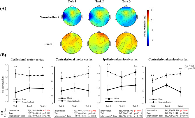
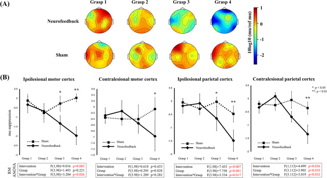
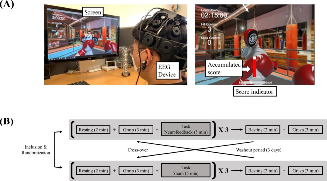
Methods: A total of 15 patients with hemiplegia following subacute ischemic stroke were prospectively enrolled in this randomized cross-over study. This study comprised two experiments: neurofeedback and sham. Each experiment included four blocks: three blocks of resting, grasp, resting, and an interventional task, followed by one block of resting and grasp. During the resting sessions, participants fixated on a white cross on a black background for 2 min without moving their upper extremities. In the grasp sessions, participants were instructed to grasp and release their paretic hand at a frequency of about 1 Hz for 3 min while maintaining fixation on the white cross. During the interventional task, the neurofeedback presented a punching image using the affected upper limb, corresponding to the mu suppression induced by imagined movement for 3 min. In contrast, the sham presented an image based on mu suppression data from randomly selected participants. EEG data were recorded throughout the experiment, and data from electrodes C3/C4 and P3/P4 were analyzed to compare the degree of mu suppression between the neurofeedback and sham experiments.
Results: Significant mu suppression was observed in the bilateral motor and parietal cortices during the neurofeedback experiment compared with the sham across serial sessions (p < 0.001). Following neurofeedback, real grasping sessions showed progressive strengthening of mu suppression in the ipsilesional motor cortex and bilateral parietal cortices compared to sessions following sham (p < 0.05). This effect was not observed in the contralesional motor cortex.
Conclusions: Motor imagery neurofeedback significantly enhances mu suppression in the ipsilesional motor and bilateral parietal cortices during motor attempts in patients with subacute stroke. These findings suggest that motor imagery neurofeedback could serve as a promising adjunctive therapy to enhance motor-related cortical activity and support motor rehabilitation in patients with stroke.
期刊介绍:
Journal of NeuroEngineering and Rehabilitation considers manuscripts on all aspects of research that result from cross-fertilization of the fields of neuroscience, biomedical engineering, and physical medicine & rehabilitation.
 求助内容:
求助内容: 应助结果提醒方式:


