基于空间模型检测的符号和混合人工智能脑组织分割
IF 6.2
2区 医学
Q1 COMPUTER SCIENCE, ARTIFICIAL INTELLIGENCE
引用次数: 0
摘要
三维医学图像的分割,特别是脑图像的分割,是神经影像学和放射治疗中的一个重要课题。因此,克服目前人工描绘脑肿瘤耗时的做法,并提供一种准确、可解释和可复制的肿瘤区域和相关组织分割方法,是一项开放的研究挑战。在本文中,我们首先提出了一种新的基于空间模型检查的脑损伤分割和描绘的符号方法。该方法的基础是闭包空间理论、拓扑空间的推广和空间逻辑。其核心是用于图像分析的高级声明性逻辑语言ImgQL和高效的空间模型检查器VoxLogicA,在其模型检查算法中利用了最先进的图像分析库。然后,我们说明了如何将该技术与机器学习技术相结合,从而形成一种混合人工智能方法,提供准确且可解释的分割结果。我们展示了符号方法在几个具有三维磁共振(MR)图像的公共数据集上的应用结果。2017年、2019年和2020年国际MICCAI BraTS挑战赛提供了三个数据集,分别包含210张、259张和293张MR图像,第四个数据集是BrainWeb数据集,包含20张(合成)正常大脑的3D患者图像。然后,我们将混合人工智能方法应用于BraTS 2020训练集。我们的分割结果显示与其他最近的方法相一致,无论是从准确性的角度还是从计算效率的角度来看,都是最先进的,但具有可解释的优势。本文章由计算机程序翻译,如有差异,请以英文原文为准。
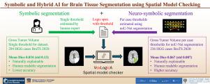
Symbolic and hybrid AI for brain tissue segmentation using spatial model checking
Segmentation of 3D medical images, and brain segmentation in particular, is an important topic in neuroimaging and in radiotherapy. Overcoming the current, time consuming, practise of manual delineation of brain tumours and providing an accurate, explainable, and replicable method of segmentation of the tumour area and related tissues is therefore an open research challenge.
In this paper, we first propose a novel symbolic approach to brain segmentation and delineation of brain lesions based on spatial model checking. This method has its foundations in the theory of closure spaces, a generalisation of topological spaces, and spatial logics. At its core is a high-level declarative logic language for image analysis, ImgQL, and an efficient spatial model checker, VoxLogicA, exploiting state-of-the-art image analysis libraries in its model checking algorithm. We then illustrate how this technique can be combined with Machine Learning techniques leading to a hybrid AI approach that provides accurate and explainable segmentation results.
We show the results of the application of the symbolic approach on several public datasets with 3D magnetic resonance (MR) images. Three datasets are provided by the 2017, 2019 and 2020 international MICCAI BraTS Challenges with 210, 259 and 293 MR images, respectively, and the fourth is the BrainWeb dataset with 20 (synthetic) 3D patient images of the normal brain. We then apply the hybrid AI method to the BraTS 2020 training set. Our segmentation results are shown to be in line with the state-of-the-art with respect to other recent approaches, both from the accuracy point of view as well as from the view of computational efficiency, but with the advantage of them being explainable.
求助全文
通过发布文献求助,成功后即可免费获取论文全文。
去求助
来源期刊
Artificial Intelligence in Medicine
工程技术-工程:生物医学
CiteScore
15.00
自引率
2.70%
发文量
143
审稿时长
6.3 months
期刊介绍:
Artificial Intelligence in Medicine publishes original articles from a wide variety of interdisciplinary perspectives concerning the theory and practice of artificial intelligence (AI) in medicine, medically-oriented human biology, and health care.
Artificial intelligence in medicine may be characterized as the scientific discipline pertaining to research studies, projects, and applications that aim at supporting decision-based medical tasks through knowledge- and/or data-intensive computer-based solutions that ultimately support and improve the performance of a human care provider.
 求助内容:
求助内容: 应助结果提醒方式:


