Carolyn Cook, Lydia Turk, Alicia January, Teresa Cappello, Peter Smith
{"title":"成骨不全患者长骨皮质透光病变的评价。","authors":"Carolyn Cook, Lydia Turk, Alicia January, Teresa Cappello, Peter Smith","doi":"10.1016/j.jposna.2024.100128","DOIUrl":null,"url":null,"abstract":"<p><strong>Background: </strong>Osteogenesis imperfecta (OI), a rare genetic disorder of collagen synthesis and metabolism, is characterized by cortical bone thinning and decreased trabecular bone. We have noted individuals with OI develop radiolucent lesions in the cortices of the long bones and these have not been previously described. The prevalence of these lesions in the long bones of children and the significance of their appearance in relation to subsequent fractures is unknown.</p><p><strong>Methods: </strong>In a retrospective study, upper and lower extremity radiographs of 328 pediatric patients with OI were examined to determine the occurrence and clinical course of these lesions in patients with various subtypes of OI.</p><p><strong>Results: </strong>Lesions were present in 55 (17%) of the 328 patients. A total of 89 lesions were identified in these 55 patients, with 138 positive radiographs across the OI subtypes. Of the 138 positive radiographs, lesions were present in 90/138 (65%) tibiae, 10/138 (7%) fibulae, 24/138 (17%) femora, 4/138 (3%) ulnae, 1/138 (1%) radius, and 9/138 (7%) humeri. In affected patients, there was an average of 1.62 lesions per patient. In 99% (136/138) of the radiographs, the lesion was present on the diaphysis of the long bone, and in 86% (119/138) of the x-rays, the lesion was located on the tension side. 64% (57/89) of the lesions developed after a previous fracture, and in 24% (21/89) of lesions, a fracture subsequently occurred through the area of the lesion.</p><p><strong>Conclusions: </strong>We present that radiolucent lesions of long bone diaphyses are a characteristic finding in OI and often presage fractures.</p><p><strong>Key concepts: </strong>(1)Radiolucent lesions are a characteristic finding of OI and have not been yet described in literature.(2)These lesions may represent a probable failure of ossification and lack of remodeling in the area of the lesion.(3)There is no clear etiology of this finding, although many arise in areas of prior fracture or osteotomy.</p>","PeriodicalId":520850,"journal":{"name":"Journal of the Pediatric Orthopaedic Society of North America","volume":"9 ","pages":"100128"},"PeriodicalIF":0.0000,"publicationDate":"2024-10-09","publicationTypes":"Journal Article","fieldsOfStudy":null,"isOpenAccess":false,"openAccessPdf":"https://www.ncbi.nlm.nih.gov/pmc/articles/PMC12088329/pdf/","citationCount":"0","resultStr":"{\"title\":\"Evaluation of Radiolucent Lesions in Cortices of Long Bones in Osteogenesis Imperfecta Patients.\",\"authors\":\"Carolyn Cook, Lydia Turk, Alicia January, Teresa Cappello, Peter Smith\",\"doi\":\"10.1016/j.jposna.2024.100128\",\"DOIUrl\":null,\"url\":null,\"abstract\":\"<p><strong>Background: </strong>Osteogenesis imperfecta (OI), a rare genetic disorder of collagen synthesis and metabolism, is characterized by cortical bone thinning and decreased trabecular bone. We have noted individuals with OI develop radiolucent lesions in the cortices of the long bones and these have not been previously described. The prevalence of these lesions in the long bones of children and the significance of their appearance in relation to subsequent fractures is unknown.</p><p><strong>Methods: </strong>In a retrospective study, upper and lower extremity radiographs of 328 pediatric patients with OI were examined to determine the occurrence and clinical course of these lesions in patients with various subtypes of OI.</p><p><strong>Results: </strong>Lesions were present in 55 (17%) of the 328 patients. A total of 89 lesions were identified in these 55 patients, with 138 positive radiographs across the OI subtypes. Of the 138 positive radiographs, lesions were present in 90/138 (65%) tibiae, 10/138 (7%) fibulae, 24/138 (17%) femora, 4/138 (3%) ulnae, 1/138 (1%) radius, and 9/138 (7%) humeri. In affected patients, there was an average of 1.62 lesions per patient. In 99% (136/138) of the radiographs, the lesion was present on the diaphysis of the long bone, and in 86% (119/138) of the x-rays, the lesion was located on the tension side. 64% (57/89) of the lesions developed after a previous fracture, and in 24% (21/89) of lesions, a fracture subsequently occurred through the area of the lesion.</p><p><strong>Conclusions: </strong>We present that radiolucent lesions of long bone diaphyses are a characteristic finding in OI and often presage fractures.</p><p><strong>Key concepts: </strong>(1)Radiolucent lesions are a characteristic finding of OI and have not been yet described in literature.(2)These lesions may represent a probable failure of ossification and lack of remodeling in the area of the lesion.(3)There is no clear etiology of this finding, although many arise in areas of prior fracture or osteotomy.</p>\",\"PeriodicalId\":520850,\"journal\":{\"name\":\"Journal of the Pediatric Orthopaedic Society of North America\",\"volume\":\"9 \",\"pages\":\"100128\"},\"PeriodicalIF\":0.0000,\"publicationDate\":\"2024-10-09\",\"publicationTypes\":\"Journal Article\",\"fieldsOfStudy\":null,\"isOpenAccess\":false,\"openAccessPdf\":\"https://www.ncbi.nlm.nih.gov/pmc/articles/PMC12088329/pdf/\",\"citationCount\":\"0\",\"resultStr\":null,\"platform\":\"Semanticscholar\",\"paperid\":null,\"PeriodicalName\":\"Journal of the Pediatric Orthopaedic Society of North America\",\"FirstCategoryId\":\"1085\",\"ListUrlMain\":\"https://doi.org/10.1016/j.jposna.2024.100128\",\"RegionNum\":0,\"RegionCategory\":null,\"ArticlePicture\":[],\"TitleCN\":null,\"AbstractTextCN\":null,\"PMCID\":null,\"EPubDate\":\"2024/11/1 0:00:00\",\"PubModel\":\"eCollection\",\"JCR\":\"\",\"JCRName\":\"\",\"Score\":null,\"Total\":0}","platform":"Semanticscholar","paperid":null,"PeriodicalName":"Journal of the Pediatric Orthopaedic Society of North America","FirstCategoryId":"1085","ListUrlMain":"https://doi.org/10.1016/j.jposna.2024.100128","RegionNum":0,"RegionCategory":null,"ArticlePicture":[],"TitleCN":null,"AbstractTextCN":null,"PMCID":null,"EPubDate":"2024/11/1 0:00:00","PubModel":"eCollection","JCR":"","JCRName":"","Score":null,"Total":0}
Evaluation of Radiolucent Lesions in Cortices of Long Bones in Osteogenesis Imperfecta Patients.
Background: Osteogenesis imperfecta (OI), a rare genetic disorder of collagen synthesis and metabolism, is characterized by cortical bone thinning and decreased trabecular bone. We have noted individuals with OI develop radiolucent lesions in the cortices of the long bones and these have not been previously described. The prevalence of these lesions in the long bones of children and the significance of their appearance in relation to subsequent fractures is unknown.
Methods: In a retrospective study, upper and lower extremity radiographs of 328 pediatric patients with OI were examined to determine the occurrence and clinical course of these lesions in patients with various subtypes of OI.
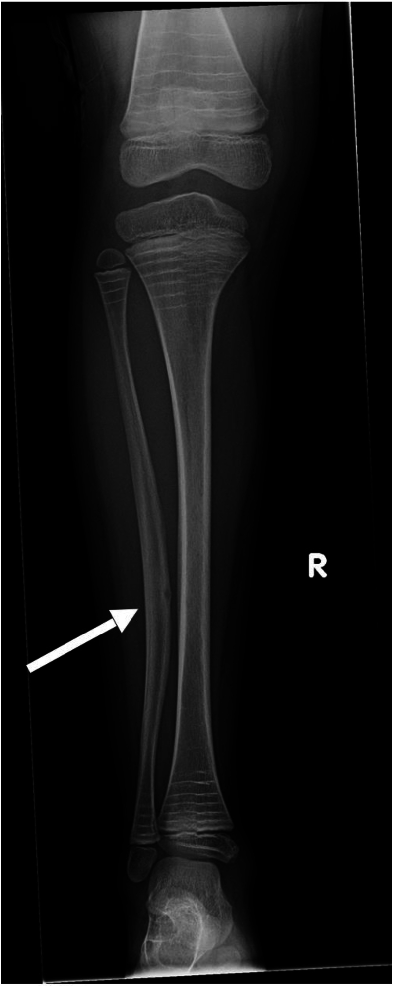
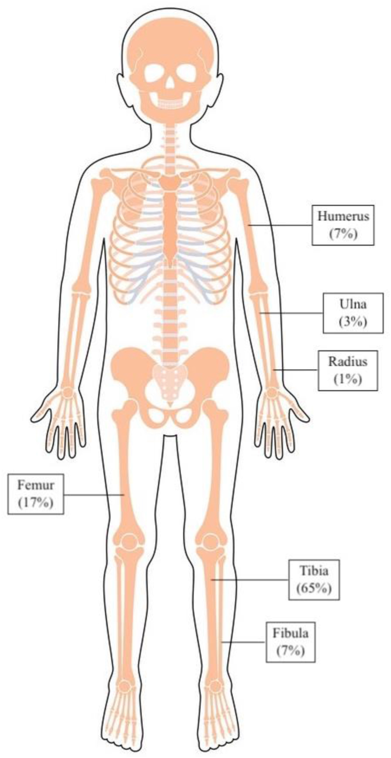
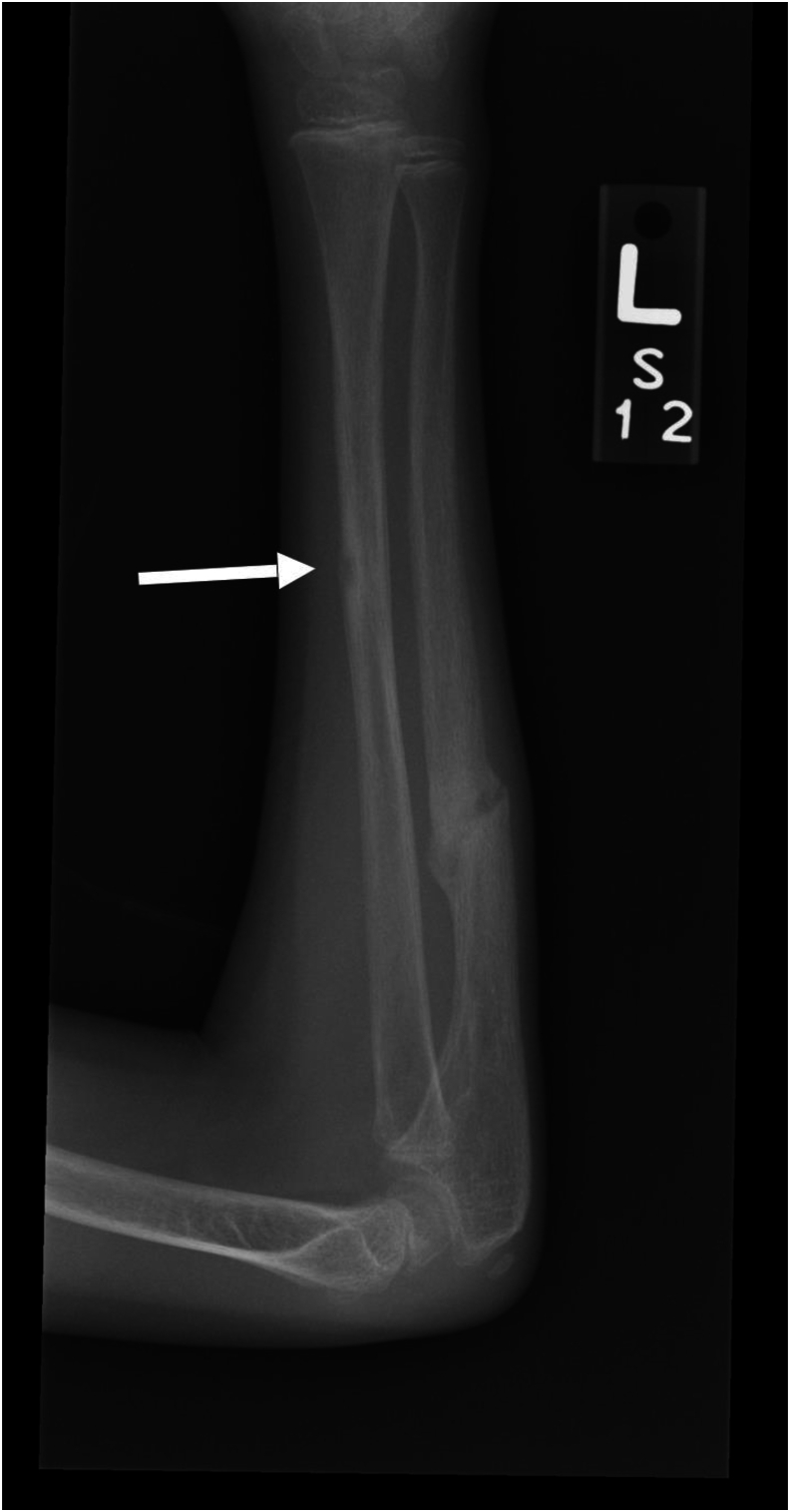
Results: Lesions were present in 55 (17%) of the 328 patients. A total of 89 lesions were identified in these 55 patients, with 138 positive radiographs across the OI subtypes. Of the 138 positive radiographs, lesions were present in 90/138 (65%) tibiae, 10/138 (7%) fibulae, 24/138 (17%) femora, 4/138 (3%) ulnae, 1/138 (1%) radius, and 9/138 (7%) humeri. In affected patients, there was an average of 1.62 lesions per patient. In 99% (136/138) of the radiographs, the lesion was present on the diaphysis of the long bone, and in 86% (119/138) of the x-rays, the lesion was located on the tension side. 64% (57/89) of the lesions developed after a previous fracture, and in 24% (21/89) of lesions, a fracture subsequently occurred through the area of the lesion.
Conclusions: We present that radiolucent lesions of long bone diaphyses are a characteristic finding in OI and often presage fractures.
Key concepts: (1)Radiolucent lesions are a characteristic finding of OI and have not been yet described in literature.(2)These lesions may represent a probable failure of ossification and lack of remodeling in the area of the lesion.(3)There is no clear etiology of this finding, although many arise in areas of prior fracture or osteotomy.

 求助内容:
求助内容: 应助结果提醒方式:


