{"title":"基于深度学习图像重建的双能谱CT图像分割性能评价:一个幻影研究。","authors":"Haoyan Li, Zhenpeng Chen, Shuaiyi Gao, Jiaqi Hu, Zhihao Yang, Yun Peng, Jihang Sun","doi":"10.3390/tomography11050051","DOIUrl":null,"url":null,"abstract":"<p><p><b>Objectives</b>: To evaluate the medical image segmentation performance of monochromatic images in various energy levels. <b>Methods</b>: The low-density module (25 mm in diameter, 6 Hounsfield Unit (HU) in density difference from background) from the ACR464 phantom was scanned at both 10 mGy and 5 mGy dose levels. Virtual monoenergetic images (VMIs) at different energy levels of 40, 50, 60, 68, 74, and 100 keV were generated. The images at 10 mGy reconstructed with 50% adaptive statistical iterative reconstruction veo (ASIR-V50%) were used to train an image segmentation model based on U-Net. The evaluation set used 5 mGy VMIs reconstructed with various reconstruction algorithms: FBP, ASIR-V50%, ASIR-V100%, deep learning image reconstruction (DLIR) with low (DLIR-L), medium (DLIR-M), and high (DLIR-H) strength levels. U-Net was employed as a tool to compare algorithm performance. Image noise and segmentation metrics, such as the DICE coefficient, intersection over union (IOU), sensitivity, and Hausdorff distance, were calculated to assess both image quality and segmentation performance. <b>Results</b>: DLIR-M and DLIR-H consistently achieved lower image noise and better segmentation performance, with the highest results observed at 60 keV, and DLIR-H had the lowest image noise across all energy levels. The performance metrics, including IOU, DICE, and sensitivity, were ranked in descending order with energy levels of 60 keV, 68 keV, 50 keV, 74 keV, 40 keV, and 100 keV. Specifically, at 60 keV, the average IOU values for each reconstruction method were 0.60 for FBP, 0.67 for ASIR-V50%, 0.68 for ASIR-V100%, 0.72 for DLIR-L, 0.75 for DLIR-M, and 0.75 for DLIR-H. The average DICE values were 0.75, 0.80, 0.82, 0.83, 0.85, and 0.86. The sensitivity values were 0.93, 0.91, 0.96, 0.95, 0.98, and 0.98. <b>Conclusions</b>: For low-density, non-enhancing objects under a low dose, the 60 keV VMIs performed better in automatic segmentation. DLIR-M and DLIR-H algorithms delivered the best results, whereas DLIR-H provided the lowest image noise and highest sensitivity.</p>","PeriodicalId":51330,"journal":{"name":"Tomography","volume":"11 5","pages":""},"PeriodicalIF":2.2000,"publicationDate":"2025-04-27","publicationTypes":"Journal Article","fieldsOfStudy":null,"isOpenAccess":false,"openAccessPdf":"https://www.ncbi.nlm.nih.gov/pmc/articles/PMC12116077/pdf/","citationCount":"0","resultStr":"{\"title\":\"Performance Evaluation of Image Segmentation Using Dual-Energy Spectral CT Images with Deep Learning Image Reconstruction: A Phantom Study.\",\"authors\":\"Haoyan Li, Zhenpeng Chen, Shuaiyi Gao, Jiaqi Hu, Zhihao Yang, Yun Peng, Jihang Sun\",\"doi\":\"10.3390/tomography11050051\",\"DOIUrl\":null,\"url\":null,\"abstract\":\"<p><p><b>Objectives</b>: To evaluate the medical image segmentation performance of monochromatic images in various energy levels. <b>Methods</b>: The low-density module (25 mm in diameter, 6 Hounsfield Unit (HU) in density difference from background) from the ACR464 phantom was scanned at both 10 mGy and 5 mGy dose levels. Virtual monoenergetic images (VMIs) at different energy levels of 40, 50, 60, 68, 74, and 100 keV were generated. The images at 10 mGy reconstructed with 50% adaptive statistical iterative reconstruction veo (ASIR-V50%) were used to train an image segmentation model based on U-Net. The evaluation set used 5 mGy VMIs reconstructed with various reconstruction algorithms: FBP, ASIR-V50%, ASIR-V100%, deep learning image reconstruction (DLIR) with low (DLIR-L), medium (DLIR-M), and high (DLIR-H) strength levels. U-Net was employed as a tool to compare algorithm performance. Image noise and segmentation metrics, such as the DICE coefficient, intersection over union (IOU), sensitivity, and Hausdorff distance, were calculated to assess both image quality and segmentation performance. <b>Results</b>: DLIR-M and DLIR-H consistently achieved lower image noise and better segmentation performance, with the highest results observed at 60 keV, and DLIR-H had the lowest image noise across all energy levels. The performance metrics, including IOU, DICE, and sensitivity, were ranked in descending order with energy levels of 60 keV, 68 keV, 50 keV, 74 keV, 40 keV, and 100 keV. Specifically, at 60 keV, the average IOU values for each reconstruction method were 0.60 for FBP, 0.67 for ASIR-V50%, 0.68 for ASIR-V100%, 0.72 for DLIR-L, 0.75 for DLIR-M, and 0.75 for DLIR-H. The average DICE values were 0.75, 0.80, 0.82, 0.83, 0.85, and 0.86. The sensitivity values were 0.93, 0.91, 0.96, 0.95, 0.98, and 0.98. <b>Conclusions</b>: For low-density, non-enhancing objects under a low dose, the 60 keV VMIs performed better in automatic segmentation. DLIR-M and DLIR-H algorithms delivered the best results, whereas DLIR-H provided the lowest image noise and highest sensitivity.</p>\",\"PeriodicalId\":51330,\"journal\":{\"name\":\"Tomography\",\"volume\":\"11 5\",\"pages\":\"\"},\"PeriodicalIF\":2.2000,\"publicationDate\":\"2025-04-27\",\"publicationTypes\":\"Journal Article\",\"fieldsOfStudy\":null,\"isOpenAccess\":false,\"openAccessPdf\":\"https://www.ncbi.nlm.nih.gov/pmc/articles/PMC12116077/pdf/\",\"citationCount\":\"0\",\"resultStr\":null,\"platform\":\"Semanticscholar\",\"paperid\":null,\"PeriodicalName\":\"Tomography\",\"FirstCategoryId\":\"3\",\"ListUrlMain\":\"https://doi.org/10.3390/tomography11050051\",\"RegionNum\":4,\"RegionCategory\":\"医学\",\"ArticlePicture\":[],\"TitleCN\":null,\"AbstractTextCN\":null,\"PMCID\":null,\"EPubDate\":\"\",\"PubModel\":\"\",\"JCR\":\"Q2\",\"JCRName\":\"RADIOLOGY, NUCLEAR MEDICINE & MEDICAL IMAGING\",\"Score\":null,\"Total\":0}","platform":"Semanticscholar","paperid":null,"PeriodicalName":"Tomography","FirstCategoryId":"3","ListUrlMain":"https://doi.org/10.3390/tomography11050051","RegionNum":4,"RegionCategory":"医学","ArticlePicture":[],"TitleCN":null,"AbstractTextCN":null,"PMCID":null,"EPubDate":"","PubModel":"","JCR":"Q2","JCRName":"RADIOLOGY, NUCLEAR MEDICINE & MEDICAL IMAGING","Score":null,"Total":0}
Performance Evaluation of Image Segmentation Using Dual-Energy Spectral CT Images with Deep Learning Image Reconstruction: A Phantom Study.
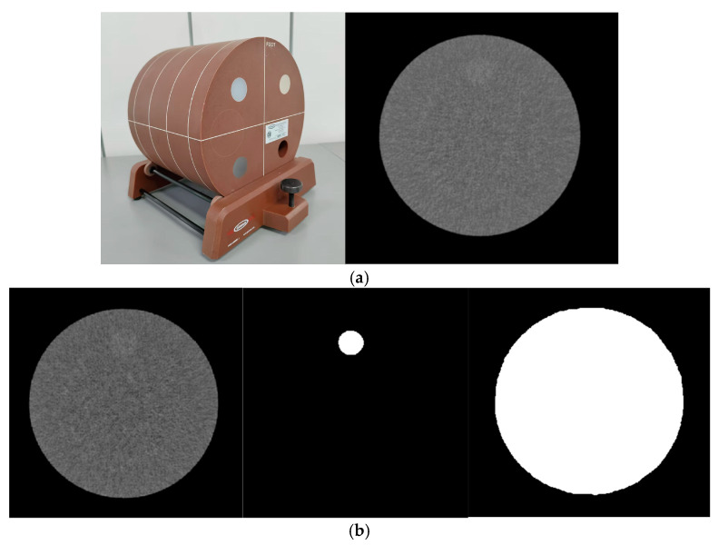
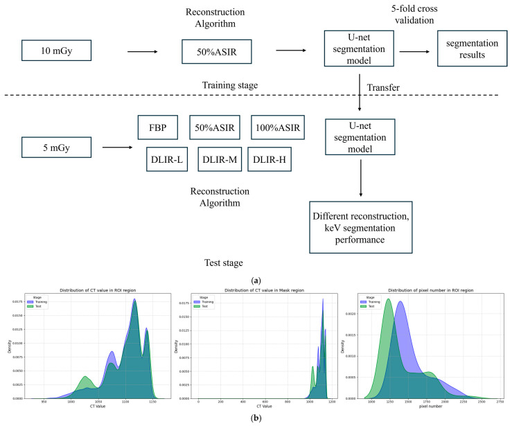
Objectives: To evaluate the medical image segmentation performance of monochromatic images in various energy levels. Methods: The low-density module (25 mm in diameter, 6 Hounsfield Unit (HU) in density difference from background) from the ACR464 phantom was scanned at both 10 mGy and 5 mGy dose levels. Virtual monoenergetic images (VMIs) at different energy levels of 40, 50, 60, 68, 74, and 100 keV were generated. The images at 10 mGy reconstructed with 50% adaptive statistical iterative reconstruction veo (ASIR-V50%) were used to train an image segmentation model based on U-Net. The evaluation set used 5 mGy VMIs reconstructed with various reconstruction algorithms: FBP, ASIR-V50%, ASIR-V100%, deep learning image reconstruction (DLIR) with low (DLIR-L), medium (DLIR-M), and high (DLIR-H) strength levels. U-Net was employed as a tool to compare algorithm performance. Image noise and segmentation metrics, such as the DICE coefficient, intersection over union (IOU), sensitivity, and Hausdorff distance, were calculated to assess both image quality and segmentation performance. Results: DLIR-M and DLIR-H consistently achieved lower image noise and better segmentation performance, with the highest results observed at 60 keV, and DLIR-H had the lowest image noise across all energy levels. The performance metrics, including IOU, DICE, and sensitivity, were ranked in descending order with energy levels of 60 keV, 68 keV, 50 keV, 74 keV, 40 keV, and 100 keV. Specifically, at 60 keV, the average IOU values for each reconstruction method were 0.60 for FBP, 0.67 for ASIR-V50%, 0.68 for ASIR-V100%, 0.72 for DLIR-L, 0.75 for DLIR-M, and 0.75 for DLIR-H. The average DICE values were 0.75, 0.80, 0.82, 0.83, 0.85, and 0.86. The sensitivity values were 0.93, 0.91, 0.96, 0.95, 0.98, and 0.98. Conclusions: For low-density, non-enhancing objects under a low dose, the 60 keV VMIs performed better in automatic segmentation. DLIR-M and DLIR-H algorithms delivered the best results, whereas DLIR-H provided the lowest image noise and highest sensitivity.
TomographyMedicine-Radiology, Nuclear Medicine and Imaging
CiteScore
2.70
自引率
10.50%
发文量
222
期刊介绍:
TomographyTM publishes basic (technical and pre-clinical) and clinical scientific articles which involve the advancement of imaging technologies. Tomography encompasses studies that use single or multiple imaging modalities including for example CT, US, PET, SPECT, MR and hyperpolarization technologies, as well as optical modalities (i.e. bioluminescence, photoacoustic, endomicroscopy, fiber optic imaging and optical computed tomography) in basic sciences, engineering, preclinical and clinical medicine.
Tomography also welcomes studies involving exploration and refinement of contrast mechanisms and image-derived metrics within and across modalities toward the development of novel imaging probes for image-based feedback and intervention. The use of imaging in biology and medicine provides unparalleled opportunities to noninvasively interrogate tissues to obtain real-time dynamic and quantitative information required for diagnosis and response to interventions and to follow evolving pathological conditions. As multi-modal studies and the complexities of imaging technologies themselves are ever increasing to provide advanced information to scientists and clinicians.
Tomography provides a unique publication venue allowing investigators the opportunity to more precisely communicate integrated findings related to the diverse and heterogeneous features associated with underlying anatomical, physiological, functional, metabolic and molecular genetic activities of normal and diseased tissue. Thus Tomography publishes peer-reviewed articles which involve the broad use of imaging of any tissue and disease type including both preclinical and clinical investigations. In addition, hardware/software along with chemical and molecular probe advances are welcome as they are deemed to significantly contribute towards the long-term goal of improving the overall impact of imaging on scientific and clinical discovery.

 求助内容:
求助内容: 应助结果提醒方式:


