Zeynep Keskin, Mihrican Yeşildağ, Ömer Özberk, Kemal Ödev, Fatih Ateş, Bengü Özkan Bakdık, Şehriban Çağlak Kardaş
{"title":"COVID-19的长期影响:2020年至2024年计算机断层扫描评估患者影像学表现分析","authors":"Zeynep Keskin, Mihrican Yeşildağ, Ömer Özberk, Kemal Ödev, Fatih Ateş, Bengü Özkan Bakdık, Şehriban Çağlak Kardaş","doi":"10.3390/tomography11050049","DOIUrl":null,"url":null,"abstract":"<p><p><b>Background:</b> This study aims to systematically evaluate the findings from computed tomography (CT) examinations conducted at least three months post-diagnosis of COVID-19 in patients diagnosed between 2020 and 2024. <b>Objective:</b> To determine the frequency and characteristics of CT findings in the post-COVID-19 period, analyze long-term effects on lung parenchyma, and contribute to the development of clinical follow-up and treatment strategies based on the collected data. <b>Materials and Methods:</b> Ethical approval was obtained for this retrospective study, and individual consent was waived. A total of 76 patients were included in the study, aged 18 and older, diagnosed with COVID-19 between March 2020 and November 2024, who underwent follow-up chest CT scans at 3-6 months, 6-12 months, and/or 12 months post-diagnosis. CT images were obtained in the supine position without contrast and evaluated by two experienced radiologists using a CT severity score (CT-SS) system, which quantifies lung involvement. Statistical analyses were performed using IBM SPSS 23.0, with significance set at <i>p</i> < 0.05. <b>Results:</b> The results indicated a mean CT-SS of 10.58 ± 0.659. Significant associations were found between age, CT scores, and the necessity for intensive care or mechanical ventilation. The most common CT findings included ground-glass opacities, reticular patterns, and traction bronchiectasis, particularly increasing with age and over time. <b>Conclusion:</b> This study emphasizes the persistent alterations in lung parenchyma following COVID-19, highlighting the importance of continuous monitoring and tailored treatment strategies for affected patients to improve long-term outcomes.</p>","PeriodicalId":51330,"journal":{"name":"Tomography","volume":"11 5","pages":""},"PeriodicalIF":2.2000,"publicationDate":"2025-04-24","publicationTypes":"Journal Article","fieldsOfStudy":null,"isOpenAccess":false,"openAccessPdf":"https://www.ncbi.nlm.nih.gov/pmc/articles/PMC12115870/pdf/","citationCount":"0","resultStr":"{\"title\":\"Long-Term Effects of COVID-19: Analysis of Imaging Findings in Patients Evaluated by Computed Tomography from 2020 to 2024.\",\"authors\":\"Zeynep Keskin, Mihrican Yeşildağ, Ömer Özberk, Kemal Ödev, Fatih Ateş, Bengü Özkan Bakdık, Şehriban Çağlak Kardaş\",\"doi\":\"10.3390/tomography11050049\",\"DOIUrl\":null,\"url\":null,\"abstract\":\"<p><p><b>Background:</b> This study aims to systematically evaluate the findings from computed tomography (CT) examinations conducted at least three months post-diagnosis of COVID-19 in patients diagnosed between 2020 and 2024. <b>Objective:</b> To determine the frequency and characteristics of CT findings in the post-COVID-19 period, analyze long-term effects on lung parenchyma, and contribute to the development of clinical follow-up and treatment strategies based on the collected data. <b>Materials and Methods:</b> Ethical approval was obtained for this retrospective study, and individual consent was waived. A total of 76 patients were included in the study, aged 18 and older, diagnosed with COVID-19 between March 2020 and November 2024, who underwent follow-up chest CT scans at 3-6 months, 6-12 months, and/or 12 months post-diagnosis. CT images were obtained in the supine position without contrast and evaluated by two experienced radiologists using a CT severity score (CT-SS) system, which quantifies lung involvement. Statistical analyses were performed using IBM SPSS 23.0, with significance set at <i>p</i> < 0.05. <b>Results:</b> The results indicated a mean CT-SS of 10.58 ± 0.659. Significant associations were found between age, CT scores, and the necessity for intensive care or mechanical ventilation. The most common CT findings included ground-glass opacities, reticular patterns, and traction bronchiectasis, particularly increasing with age and over time. <b>Conclusion:</b> This study emphasizes the persistent alterations in lung parenchyma following COVID-19, highlighting the importance of continuous monitoring and tailored treatment strategies for affected patients to improve long-term outcomes.</p>\",\"PeriodicalId\":51330,\"journal\":{\"name\":\"Tomography\",\"volume\":\"11 5\",\"pages\":\"\"},\"PeriodicalIF\":2.2000,\"publicationDate\":\"2025-04-24\",\"publicationTypes\":\"Journal Article\",\"fieldsOfStudy\":null,\"isOpenAccess\":false,\"openAccessPdf\":\"https://www.ncbi.nlm.nih.gov/pmc/articles/PMC12115870/pdf/\",\"citationCount\":\"0\",\"resultStr\":null,\"platform\":\"Semanticscholar\",\"paperid\":null,\"PeriodicalName\":\"Tomography\",\"FirstCategoryId\":\"3\",\"ListUrlMain\":\"https://doi.org/10.3390/tomography11050049\",\"RegionNum\":4,\"RegionCategory\":\"医学\",\"ArticlePicture\":[],\"TitleCN\":null,\"AbstractTextCN\":null,\"PMCID\":null,\"EPubDate\":\"\",\"PubModel\":\"\",\"JCR\":\"Q2\",\"JCRName\":\"RADIOLOGY, NUCLEAR MEDICINE & MEDICAL IMAGING\",\"Score\":null,\"Total\":0}","platform":"Semanticscholar","paperid":null,"PeriodicalName":"Tomography","FirstCategoryId":"3","ListUrlMain":"https://doi.org/10.3390/tomography11050049","RegionNum":4,"RegionCategory":"医学","ArticlePicture":[],"TitleCN":null,"AbstractTextCN":null,"PMCID":null,"EPubDate":"","PubModel":"","JCR":"Q2","JCRName":"RADIOLOGY, NUCLEAR MEDICINE & MEDICAL IMAGING","Score":null,"Total":0}
Long-Term Effects of COVID-19: Analysis of Imaging Findings in Patients Evaluated by Computed Tomography from 2020 to 2024.
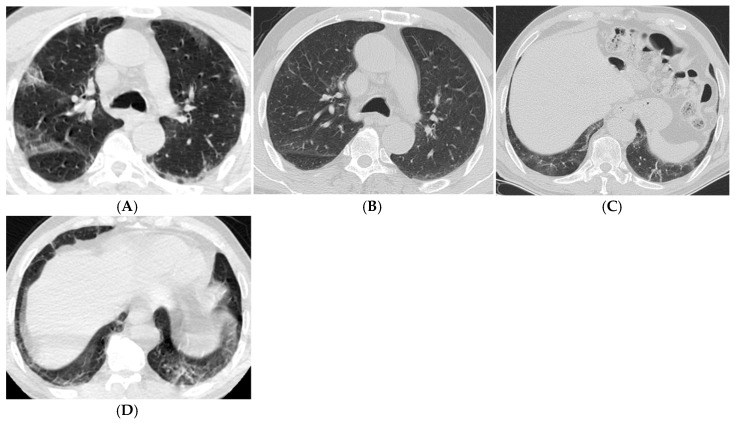
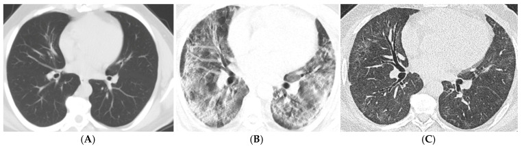
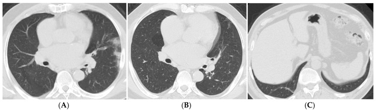
Background: This study aims to systematically evaluate the findings from computed tomography (CT) examinations conducted at least three months post-diagnosis of COVID-19 in patients diagnosed between 2020 and 2024. Objective: To determine the frequency and characteristics of CT findings in the post-COVID-19 period, analyze long-term effects on lung parenchyma, and contribute to the development of clinical follow-up and treatment strategies based on the collected data. Materials and Methods: Ethical approval was obtained for this retrospective study, and individual consent was waived. A total of 76 patients were included in the study, aged 18 and older, diagnosed with COVID-19 between March 2020 and November 2024, who underwent follow-up chest CT scans at 3-6 months, 6-12 months, and/or 12 months post-diagnosis. CT images were obtained in the supine position without contrast and evaluated by two experienced radiologists using a CT severity score (CT-SS) system, which quantifies lung involvement. Statistical analyses were performed using IBM SPSS 23.0, with significance set at p < 0.05. Results: The results indicated a mean CT-SS of 10.58 ± 0.659. Significant associations were found between age, CT scores, and the necessity for intensive care or mechanical ventilation. The most common CT findings included ground-glass opacities, reticular patterns, and traction bronchiectasis, particularly increasing with age and over time. Conclusion: This study emphasizes the persistent alterations in lung parenchyma following COVID-19, highlighting the importance of continuous monitoring and tailored treatment strategies for affected patients to improve long-term outcomes.
TomographyMedicine-Radiology, Nuclear Medicine and Imaging
CiteScore
2.70
自引率
10.50%
发文量
222
期刊介绍:
TomographyTM publishes basic (technical and pre-clinical) and clinical scientific articles which involve the advancement of imaging technologies. Tomography encompasses studies that use single or multiple imaging modalities including for example CT, US, PET, SPECT, MR and hyperpolarization technologies, as well as optical modalities (i.e. bioluminescence, photoacoustic, endomicroscopy, fiber optic imaging and optical computed tomography) in basic sciences, engineering, preclinical and clinical medicine.
Tomography also welcomes studies involving exploration and refinement of contrast mechanisms and image-derived metrics within and across modalities toward the development of novel imaging probes for image-based feedback and intervention. The use of imaging in biology and medicine provides unparalleled opportunities to noninvasively interrogate tissues to obtain real-time dynamic and quantitative information required for diagnosis and response to interventions and to follow evolving pathological conditions. As multi-modal studies and the complexities of imaging technologies themselves are ever increasing to provide advanced information to scientists and clinicians.
Tomography provides a unique publication venue allowing investigators the opportunity to more precisely communicate integrated findings related to the diverse and heterogeneous features associated with underlying anatomical, physiological, functional, metabolic and molecular genetic activities of normal and diseased tissue. Thus Tomography publishes peer-reviewed articles which involve the broad use of imaging of any tissue and disease type including both preclinical and clinical investigations. In addition, hardware/software along with chemical and molecular probe advances are welcome as they are deemed to significantly contribute towards the long-term goal of improving the overall impact of imaging on scientific and clinical discovery.

 求助内容:
求助内容: 应助结果提醒方式:


