Celal Kaya, Pınar Kendigelen, Ayşe Çiğdem Tütüncü, Güner Kaya
{"title":"评估儿童尾侧硬膜外解剖:触诊与超声鉴别骶骨角的比较。","authors":"Celal Kaya, Pınar Kendigelen, Ayşe Çiğdem Tütüncü, Güner Kaya","doi":"10.4274/TJAR.2025.251980","DOIUrl":null,"url":null,"abstract":"<p><strong>Objective: </strong>The aim of this study is to compare the identification of the sacral cornua using palpation and ultrasound, and to evaluate the sacrococcygeal area via ultrasound across different age groups of children.</p><p><strong>Methods: </strong>This study included 348 children aged 1 to 84 months, who were divided into three age groups: 1-24 months, 25-48 months, and 49-84 months. Sacral cornua were assessed using both palpation and ultrasound imaging. Palpation findings were categorized as \"good\", \"difficult\", or \"non-palpable\". Ultrasound imaging of the sacral cornua was classified as \"clear\", \"unclear\", or \"invisible\". Measurements taken included the inter-cornual distance, the anteroposterior diameter of the sacral canal, the distance from the skin to the sacral canal, and the distance from the dural sac to the cornua level.</p><p><strong>Results: </strong>Palpation of the sacral cornua was rated as \"good\" in 75.9% of patients, \"difficult\" in 22.4%, and \"non-palpable\" in 1.7%. All patients with \"good\" cornua palpation were also classified as \"clear\" on ultrasound imaging. Among the cases with \"difficult\" palpation, 76% showed a \"clear\" ultrasound image, while 24% were \"unclear\". Only one patient had \"invisible\" cornua on ultrasound. The mean distance from the dural sac to the cornua level was 3.72±1.64 cm, and this distance increased significantly with age (<i>P</i> < 0.01).</p><p><strong>Conclusion: </strong>Ultrasound is a valuable tool for identifying the sacral cornua, especially when palpation is difficult, and offers reliable, detailed information on sacral anatomy.</p>","PeriodicalId":23353,"journal":{"name":"Turkish journal of anaesthesiology and reanimation","volume":" ","pages":"107-113"},"PeriodicalIF":0.9000,"publicationDate":"2025-05-30","publicationTypes":"Journal Article","fieldsOfStudy":null,"isOpenAccess":false,"openAccessPdf":"https://www.ncbi.nlm.nih.gov/pmc/articles/PMC12123656/pdf/","citationCount":"0","resultStr":"{\"title\":\"Assessing Caudal Epidural Anatomy in Children: A Comparison of Palpation and Ultrasound for Sacral Cornua Identification.\",\"authors\":\"Celal Kaya, Pınar Kendigelen, Ayşe Çiğdem Tütüncü, Güner Kaya\",\"doi\":\"10.4274/TJAR.2025.251980\",\"DOIUrl\":null,\"url\":null,\"abstract\":\"<p><strong>Objective: </strong>The aim of this study is to compare the identification of the sacral cornua using palpation and ultrasound, and to evaluate the sacrococcygeal area via ultrasound across different age groups of children.</p><p><strong>Methods: </strong>This study included 348 children aged 1 to 84 months, who were divided into three age groups: 1-24 months, 25-48 months, and 49-84 months. Sacral cornua were assessed using both palpation and ultrasound imaging. Palpation findings were categorized as \\\"good\\\", \\\"difficult\\\", or \\\"non-palpable\\\". Ultrasound imaging of the sacral cornua was classified as \\\"clear\\\", \\\"unclear\\\", or \\\"invisible\\\". Measurements taken included the inter-cornual distance, the anteroposterior diameter of the sacral canal, the distance from the skin to the sacral canal, and the distance from the dural sac to the cornua level.</p><p><strong>Results: </strong>Palpation of the sacral cornua was rated as \\\"good\\\" in 75.9% of patients, \\\"difficult\\\" in 22.4%, and \\\"non-palpable\\\" in 1.7%. All patients with \\\"good\\\" cornua palpation were also classified as \\\"clear\\\" on ultrasound imaging. Among the cases with \\\"difficult\\\" palpation, 76% showed a \\\"clear\\\" ultrasound image, while 24% were \\\"unclear\\\". Only one patient had \\\"invisible\\\" cornua on ultrasound. The mean distance from the dural sac to the cornua level was 3.72±1.64 cm, and this distance increased significantly with age (<i>P</i> < 0.01).</p><p><strong>Conclusion: </strong>Ultrasound is a valuable tool for identifying the sacral cornua, especially when palpation is difficult, and offers reliable, detailed information on sacral anatomy.</p>\",\"PeriodicalId\":23353,\"journal\":{\"name\":\"Turkish journal of anaesthesiology and reanimation\",\"volume\":\" \",\"pages\":\"107-113\"},\"PeriodicalIF\":0.9000,\"publicationDate\":\"2025-05-30\",\"publicationTypes\":\"Journal Article\",\"fieldsOfStudy\":null,\"isOpenAccess\":false,\"openAccessPdf\":\"https://www.ncbi.nlm.nih.gov/pmc/articles/PMC12123656/pdf/\",\"citationCount\":\"0\",\"resultStr\":null,\"platform\":\"Semanticscholar\",\"paperid\":null,\"PeriodicalName\":\"Turkish journal of anaesthesiology and reanimation\",\"FirstCategoryId\":\"1085\",\"ListUrlMain\":\"https://doi.org/10.4274/TJAR.2025.251980\",\"RegionNum\":0,\"RegionCategory\":null,\"ArticlePicture\":[],\"TitleCN\":null,\"AbstractTextCN\":null,\"PMCID\":null,\"EPubDate\":\"2025/5/27 0:00:00\",\"PubModel\":\"Epub\",\"JCR\":\"Q3\",\"JCRName\":\"ANESTHESIOLOGY\",\"Score\":null,\"Total\":0}","platform":"Semanticscholar","paperid":null,"PeriodicalName":"Turkish journal of anaesthesiology and reanimation","FirstCategoryId":"1085","ListUrlMain":"https://doi.org/10.4274/TJAR.2025.251980","RegionNum":0,"RegionCategory":null,"ArticlePicture":[],"TitleCN":null,"AbstractTextCN":null,"PMCID":null,"EPubDate":"2025/5/27 0:00:00","PubModel":"Epub","JCR":"Q3","JCRName":"ANESTHESIOLOGY","Score":null,"Total":0}
Assessing Caudal Epidural Anatomy in Children: A Comparison of Palpation and Ultrasound for Sacral Cornua Identification.
Objective: The aim of this study is to compare the identification of the sacral cornua using palpation and ultrasound, and to evaluate the sacrococcygeal area via ultrasound across different age groups of children.
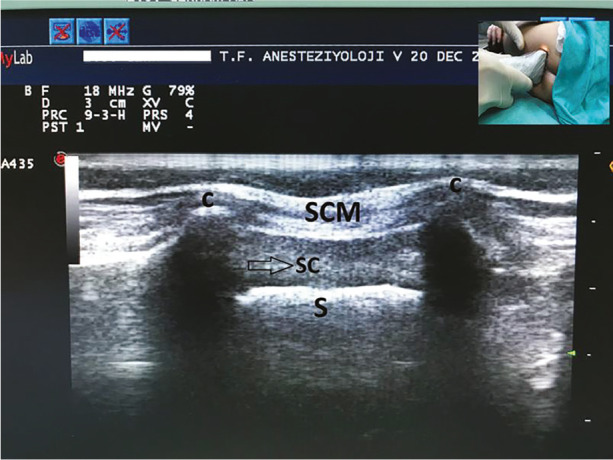
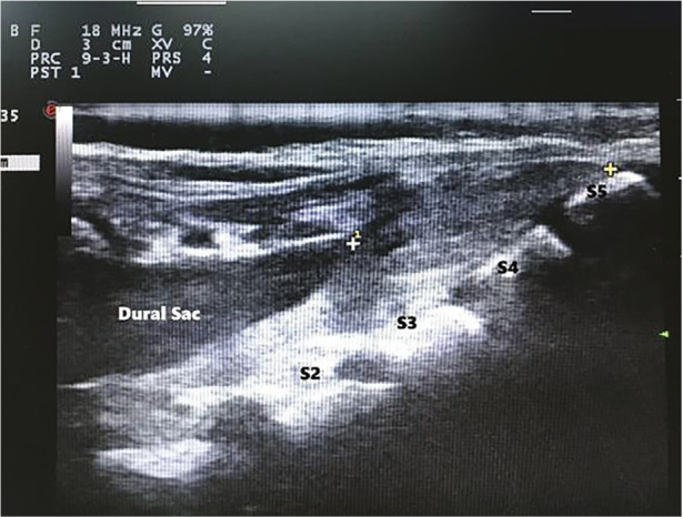
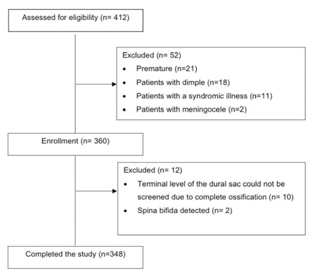
Methods: This study included 348 children aged 1 to 84 months, who were divided into three age groups: 1-24 months, 25-48 months, and 49-84 months. Sacral cornua were assessed using both palpation and ultrasound imaging. Palpation findings were categorized as "good", "difficult", or "non-palpable". Ultrasound imaging of the sacral cornua was classified as "clear", "unclear", or "invisible". Measurements taken included the inter-cornual distance, the anteroposterior diameter of the sacral canal, the distance from the skin to the sacral canal, and the distance from the dural sac to the cornua level.
Results: Palpation of the sacral cornua was rated as "good" in 75.9% of patients, "difficult" in 22.4%, and "non-palpable" in 1.7%. All patients with "good" cornua palpation were also classified as "clear" on ultrasound imaging. Among the cases with "difficult" palpation, 76% showed a "clear" ultrasound image, while 24% were "unclear". Only one patient had "invisible" cornua on ultrasound. The mean distance from the dural sac to the cornua level was 3.72±1.64 cm, and this distance increased significantly with age (P < 0.01).
Conclusion: Ultrasound is a valuable tool for identifying the sacral cornua, especially when palpation is difficult, and offers reliable, detailed information on sacral anatomy.

 求助内容:
求助内容: 应助结果提醒方式:


