{"title":"成人脑内血肿清除后出现症状性扩大的脑孔囊肿1例报告。","authors":"Yoshiaki Takamura","doi":"10.31662/jmaj.2024-0189","DOIUrl":null,"url":null,"abstract":"<p><p>Porencephalic cysts are very rare in adults. Herein, we present a case of an 88-year-old man with a symptomatic expanding porencephalic cyst after intracerebral hematoma evacuation. He was admitted because of disturbed consciousness and right hemiparesis. A computed tomography (CT) showed a large subcortical hematoma in the left parietal lobe. Hematoma evacuation was performed, his consciousness level improved but gradually deteriorated. Follow-up CT revealed a new cystic lesion with perifocal edema at the hematoma site, with progressive expansion of the cyst. Cyst drainage and -peritoneal shunt placement were performed on postoperative day 14; consequently, his symptoms improved. Considerably, a porencephalic cyst have developed because the cerebrospinal fluid flowed into the closed hematoma cavity from the ventricle owing to the osmotic pressure difference between the ventricle and the hematoma cavity.</p>","PeriodicalId":73550,"journal":{"name":"JMA journal","volume":"8 2","pages":"637-640"},"PeriodicalIF":1.8000,"publicationDate":"2025-04-28","publicationTypes":"Journal Article","fieldsOfStudy":null,"isOpenAccess":false,"openAccessPdf":"https://www.ncbi.nlm.nih.gov/pmc/articles/PMC12095862/pdf/","citationCount":"0","resultStr":"{\"title\":\"Symptomatic Expanding Porencephalic Cyst after Evacuation of Intracerebral Hematoma in an Adult: A Case Report.\",\"authors\":\"Yoshiaki Takamura\",\"doi\":\"10.31662/jmaj.2024-0189\",\"DOIUrl\":null,\"url\":null,\"abstract\":\"<p><p>Porencephalic cysts are very rare in adults. Herein, we present a case of an 88-year-old man with a symptomatic expanding porencephalic cyst after intracerebral hematoma evacuation. He was admitted because of disturbed consciousness and right hemiparesis. A computed tomography (CT) showed a large subcortical hematoma in the left parietal lobe. Hematoma evacuation was performed, his consciousness level improved but gradually deteriorated. Follow-up CT revealed a new cystic lesion with perifocal edema at the hematoma site, with progressive expansion of the cyst. Cyst drainage and -peritoneal shunt placement were performed on postoperative day 14; consequently, his symptoms improved. Considerably, a porencephalic cyst have developed because the cerebrospinal fluid flowed into the closed hematoma cavity from the ventricle owing to the osmotic pressure difference between the ventricle and the hematoma cavity.</p>\",\"PeriodicalId\":73550,\"journal\":{\"name\":\"JMA journal\",\"volume\":\"8 2\",\"pages\":\"637-640\"},\"PeriodicalIF\":1.8000,\"publicationDate\":\"2025-04-28\",\"publicationTypes\":\"Journal Article\",\"fieldsOfStudy\":null,\"isOpenAccess\":false,\"openAccessPdf\":\"https://www.ncbi.nlm.nih.gov/pmc/articles/PMC12095862/pdf/\",\"citationCount\":\"0\",\"resultStr\":null,\"platform\":\"Semanticscholar\",\"paperid\":null,\"PeriodicalName\":\"JMA journal\",\"FirstCategoryId\":\"1085\",\"ListUrlMain\":\"https://doi.org/10.31662/jmaj.2024-0189\",\"RegionNum\":0,\"RegionCategory\":null,\"ArticlePicture\":[],\"TitleCN\":null,\"AbstractTextCN\":null,\"PMCID\":null,\"EPubDate\":\"2025/2/7 0:00:00\",\"PubModel\":\"Epub\",\"JCR\":\"Q2\",\"JCRName\":\"MEDICINE, GENERAL & INTERNAL\",\"Score\":null,\"Total\":0}","platform":"Semanticscholar","paperid":null,"PeriodicalName":"JMA journal","FirstCategoryId":"1085","ListUrlMain":"https://doi.org/10.31662/jmaj.2024-0189","RegionNum":0,"RegionCategory":null,"ArticlePicture":[],"TitleCN":null,"AbstractTextCN":null,"PMCID":null,"EPubDate":"2025/2/7 0:00:00","PubModel":"Epub","JCR":"Q2","JCRName":"MEDICINE, GENERAL & INTERNAL","Score":null,"Total":0}
Symptomatic Expanding Porencephalic Cyst after Evacuation of Intracerebral Hematoma in an Adult: A Case Report.
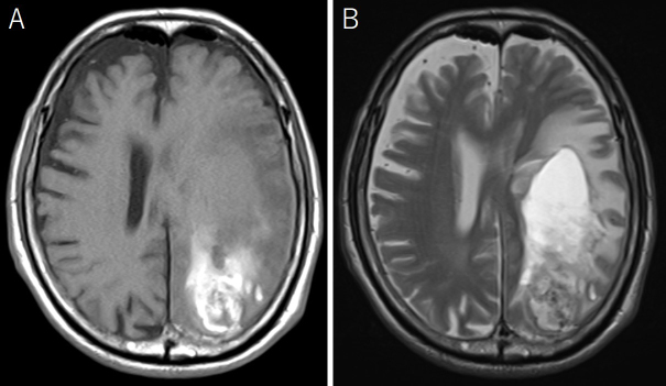
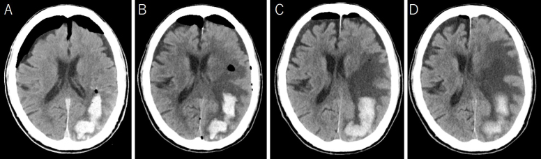
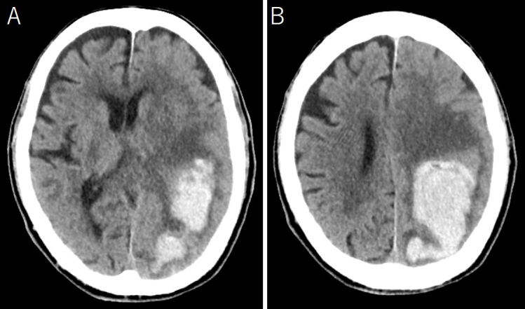
Porencephalic cysts are very rare in adults. Herein, we present a case of an 88-year-old man with a symptomatic expanding porencephalic cyst after intracerebral hematoma evacuation. He was admitted because of disturbed consciousness and right hemiparesis. A computed tomography (CT) showed a large subcortical hematoma in the left parietal lobe. Hematoma evacuation was performed, his consciousness level improved but gradually deteriorated. Follow-up CT revealed a new cystic lesion with perifocal edema at the hematoma site, with progressive expansion of the cyst. Cyst drainage and -peritoneal shunt placement were performed on postoperative day 14; consequently, his symptoms improved. Considerably, a porencephalic cyst have developed because the cerebrospinal fluid flowed into the closed hematoma cavity from the ventricle owing to the osmotic pressure difference between the ventricle and the hematoma cavity.

 求助内容:
求助内容: 应助结果提醒方式:


