{"title":"体外受精小鼠胚胎氧化应激反应激活的蛋白质组学和代谢组学研究。","authors":"Seok Hee Lee, Saúl Lira-Albarrán, Paolo F Rinaudo","doi":"10.1093/hropen/hoaf022","DOIUrl":null,"url":null,"abstract":"<p><strong>Study question: </strong>How different is the global proteomic and metabolic profile of mouse blastocysts generated by IVF, cultured in optimal (5% O<sub>2</sub>) or stressful (20% O<sub>2</sub>) conditions, compared to <i>in vivo</i> generated blastocysts?</p><p><strong>Summary answer: </strong>We found that in IVF-generated embryos: (i) the proteome was more sensitive to high oxygen levels than the global metabolomic profile; (ii) enzymes involved in splicing and the spliceosome are altered; (iii) numerous metabolic pathways, particularly amino acids metabolism, are altered (iv) there is activation of the integrated stress response (ISR) and downregulation of mTOR pathways.</p><p><strong>What is known already: </strong>IVF culture conditions are known to affect the gene expression of embryos. However, comprehensive data on the global metabolic and proteomic changes that occur in IVF-generated embryos are unknown.</p><p><strong>Study design size duration: </strong>Mouse embryos were generated by natural mating (<i>in vivo</i> control or flushed blastocyst-FB-group) or by IVF using KSOM medium and two distinct oxygen concentrations: 5% O<sub>2</sub> (optimal) and 20% O<sub>2</sub> (stressful). Proteomic and metabolomic analyses were performed using state-of-the-art mass spectrometry techniques in triplicate (n = 100 blastocysts per replicate), allowing for detailed profiling of protein and metabolite alterations in each group.</p><p><strong>Participants/materials setting methods: </strong>Mouse blastocysts were collected from CD-1 and B6D2F1 strains as specified above. High-resolution liquid chromatography-tandem mass spectrometry (LC-MS/MS) was used for proteomics, while high-performance liquid chromatography coupled with mass spectrometry (HILIC-MS) was used for metabolomics. In addition, Immunofluorescence was used to assess the activation of stress response pathways, including the ISR.</p><p><strong>Main results and the role of chance: </strong>Proteomic analysis revealed significant changes in protein expression in embryos cultured under 20% O<sub>2</sub> compared to 5% O<sub>2</sub> and <i>in vivo</i> embryos. Compared to <i>in vivo</i> embryos, IVF embryos cultured under 20% O<sub>2</sub> exhibited 599 differentially expressed proteins, with an increase in proteins involved in oxidative stress responses, aminoacyl-tRNA synthesis, and spliceosome pathways. In contrast, IVF embryos cultured under 5% O<sub>2</sub> showed fewer changes, with 426 differentially expressed proteins, though still reflecting significant alterations compared to <i>in vivo</i> embryos. These results indicate that embryos in stressful conditions (20% O<sub>2</sub>) exhibit a stronger stress response and alterations in critical pathways for protein synthesis and DNA repair. Metabolomic analysis revealed that embryos cultured under 20% O<sub>2</sub> showed changes in branch-chained amino acid levels, and decreased levels of key metabolites of the TCA cycle and pentose phosphate pathway. Embryos cultured under 5% O<sub>2</sub> had increased pyruvate levels, suggesting altered glycolysis. Immunofluorescence confirmed that oxidative stress markers such as GCN2, EIF2α, and ATF4 were upregulated in IVF embryos, indicating ISR activation. Overall, IVF and embryo culture have a direct impact on embryo proteomes and metabolomes affecting amino acid metabolism and stress-related pathways.</p><p><strong>Large scale data: </strong>N/A.</p><p><strong>Limitations reasons for caution: </strong>Results in a murine model should be extrapolated with caution to human embryos.</p><p><strong>Wider implications of the findings: </strong>These findings offer valuable insights into how different IVF culture conditions, specifically oxygen levels, impact the global metabolic and proteomic profiles of embryos. These findings provide critical insights into the profound impact of IVF culture conditions, particularly oxygen levels, on the global metabolic and proteomic landscapes of embryos. By identifying key metabolic pathways disrupted by oxidative stress, we highlight the potential clinical importance of proteomic and metabolomic analyses in understanding embryo quality, improving ART, and ultimately enhancing pregnancy outcomes. The integration of metabolomic and proteomic data offers a comprehensive understanding of how oxidative stress influences cellular function. These insights have direct clinical relevance, providing a foundation for optimizing ART protocols to mitigate oxidative stress.</p><p><strong>Study funding/competing interests: </strong>This work was supported by grant R01 HD108166-01A1 from the National Institute of Child Health and Human Development (NICHD) to P.F.R. The authors declare that there is no conflict of interest that could be perceived as prejudicing the impartiality of the research reported.</p>","PeriodicalId":73264,"journal":{"name":"Human reproduction open","volume":"2025 2","pages":"hoaf022"},"PeriodicalIF":11.1000,"publicationDate":"2025-04-28","publicationTypes":"Journal Article","fieldsOfStudy":null,"isOpenAccess":false,"openAccessPdf":"https://www.ncbi.nlm.nih.gov/pmc/articles/PMC12101870/pdf/","citationCount":"0","resultStr":"{\"title\":\"Proteomic and metabolomic insights into oxidative stress response activation in mouse embryos generated by <i>in vitro</i> fertilization.\",\"authors\":\"Seok Hee Lee, Saúl Lira-Albarrán, Paolo F Rinaudo\",\"doi\":\"10.1093/hropen/hoaf022\",\"DOIUrl\":null,\"url\":null,\"abstract\":\"<p><strong>Study question: </strong>How different is the global proteomic and metabolic profile of mouse blastocysts generated by IVF, cultured in optimal (5% O<sub>2</sub>) or stressful (20% O<sub>2</sub>) conditions, compared to <i>in vivo</i> generated blastocysts?</p><p><strong>Summary answer: </strong>We found that in IVF-generated embryos: (i) the proteome was more sensitive to high oxygen levels than the global metabolomic profile; (ii) enzymes involved in splicing and the spliceosome are altered; (iii) numerous metabolic pathways, particularly amino acids metabolism, are altered (iv) there is activation of the integrated stress response (ISR) and downregulation of mTOR pathways.</p><p><strong>What is known already: </strong>IVF culture conditions are known to affect the gene expression of embryos. However, comprehensive data on the global metabolic and proteomic changes that occur in IVF-generated embryos are unknown.</p><p><strong>Study design size duration: </strong>Mouse embryos were generated by natural mating (<i>in vivo</i> control or flushed blastocyst-FB-group) or by IVF using KSOM medium and two distinct oxygen concentrations: 5% O<sub>2</sub> (optimal) and 20% O<sub>2</sub> (stressful). Proteomic and metabolomic analyses were performed using state-of-the-art mass spectrometry techniques in triplicate (n = 100 blastocysts per replicate), allowing for detailed profiling of protein and metabolite alterations in each group.</p><p><strong>Participants/materials setting methods: </strong>Mouse blastocysts were collected from CD-1 and B6D2F1 strains as specified above. High-resolution liquid chromatography-tandem mass spectrometry (LC-MS/MS) was used for proteomics, while high-performance liquid chromatography coupled with mass spectrometry (HILIC-MS) was used for metabolomics. In addition, Immunofluorescence was used to assess the activation of stress response pathways, including the ISR.</p><p><strong>Main results and the role of chance: </strong>Proteomic analysis revealed significant changes in protein expression in embryos cultured under 20% O<sub>2</sub> compared to 5% O<sub>2</sub> and <i>in vivo</i> embryos. Compared to <i>in vivo</i> embryos, IVF embryos cultured under 20% O<sub>2</sub> exhibited 599 differentially expressed proteins, with an increase in proteins involved in oxidative stress responses, aminoacyl-tRNA synthesis, and spliceosome pathways. In contrast, IVF embryos cultured under 5% O<sub>2</sub> showed fewer changes, with 426 differentially expressed proteins, though still reflecting significant alterations compared to <i>in vivo</i> embryos. These results indicate that embryos in stressful conditions (20% O<sub>2</sub>) exhibit a stronger stress response and alterations in critical pathways for protein synthesis and DNA repair. Metabolomic analysis revealed that embryos cultured under 20% O<sub>2</sub> showed changes in branch-chained amino acid levels, and decreased levels of key metabolites of the TCA cycle and pentose phosphate pathway. Embryos cultured under 5% O<sub>2</sub> had increased pyruvate levels, suggesting altered glycolysis. Immunofluorescence confirmed that oxidative stress markers such as GCN2, EIF2α, and ATF4 were upregulated in IVF embryos, indicating ISR activation. Overall, IVF and embryo culture have a direct impact on embryo proteomes and metabolomes affecting amino acid metabolism and stress-related pathways.</p><p><strong>Large scale data: </strong>N/A.</p><p><strong>Limitations reasons for caution: </strong>Results in a murine model should be extrapolated with caution to human embryos.</p><p><strong>Wider implications of the findings: </strong>These findings offer valuable insights into how different IVF culture conditions, specifically oxygen levels, impact the global metabolic and proteomic profiles of embryos. These findings provide critical insights into the profound impact of IVF culture conditions, particularly oxygen levels, on the global metabolic and proteomic landscapes of embryos. By identifying key metabolic pathways disrupted by oxidative stress, we highlight the potential clinical importance of proteomic and metabolomic analyses in understanding embryo quality, improving ART, and ultimately enhancing pregnancy outcomes. The integration of metabolomic and proteomic data offers a comprehensive understanding of how oxidative stress influences cellular function. These insights have direct clinical relevance, providing a foundation for optimizing ART protocols to mitigate oxidative stress.</p><p><strong>Study funding/competing interests: </strong>This work was supported by grant R01 HD108166-01A1 from the National Institute of Child Health and Human Development (NICHD) to P.F.R. The authors declare that there is no conflict of interest that could be perceived as prejudicing the impartiality of the research reported.</p>\",\"PeriodicalId\":73264,\"journal\":{\"name\":\"Human reproduction open\",\"volume\":\"2025 2\",\"pages\":\"hoaf022\"},\"PeriodicalIF\":11.1000,\"publicationDate\":\"2025-04-28\",\"publicationTypes\":\"Journal Article\",\"fieldsOfStudy\":null,\"isOpenAccess\":false,\"openAccessPdf\":\"https://www.ncbi.nlm.nih.gov/pmc/articles/PMC12101870/pdf/\",\"citationCount\":\"0\",\"resultStr\":null,\"platform\":\"Semanticscholar\",\"paperid\":null,\"PeriodicalName\":\"Human reproduction open\",\"FirstCategoryId\":\"1085\",\"ListUrlMain\":\"https://doi.org/10.1093/hropen/hoaf022\",\"RegionNum\":0,\"RegionCategory\":null,\"ArticlePicture\":[],\"TitleCN\":null,\"AbstractTextCN\":null,\"PMCID\":null,\"EPubDate\":\"2025/1/1 0:00:00\",\"PubModel\":\"eCollection\",\"JCR\":\"Q1\",\"JCRName\":\"OBSTETRICS & GYNECOLOGY\",\"Score\":null,\"Total\":0}","platform":"Semanticscholar","paperid":null,"PeriodicalName":"Human reproduction open","FirstCategoryId":"1085","ListUrlMain":"https://doi.org/10.1093/hropen/hoaf022","RegionNum":0,"RegionCategory":null,"ArticlePicture":[],"TitleCN":null,"AbstractTextCN":null,"PMCID":null,"EPubDate":"2025/1/1 0:00:00","PubModel":"eCollection","JCR":"Q1","JCRName":"OBSTETRICS & GYNECOLOGY","Score":null,"Total":0}
Proteomic and metabolomic insights into oxidative stress response activation in mouse embryos generated by in vitro fertilization.
Study question: How different is the global proteomic and metabolic profile of mouse blastocysts generated by IVF, cultured in optimal (5% O2) or stressful (20% O2) conditions, compared to in vivo generated blastocysts?
Summary answer: We found that in IVF-generated embryos: (i) the proteome was more sensitive to high oxygen levels than the global metabolomic profile; (ii) enzymes involved in splicing and the spliceosome are altered; (iii) numerous metabolic pathways, particularly amino acids metabolism, are altered (iv) there is activation of the integrated stress response (ISR) and downregulation of mTOR pathways.
What is known already: IVF culture conditions are known to affect the gene expression of embryos. However, comprehensive data on the global metabolic and proteomic changes that occur in IVF-generated embryos are unknown.
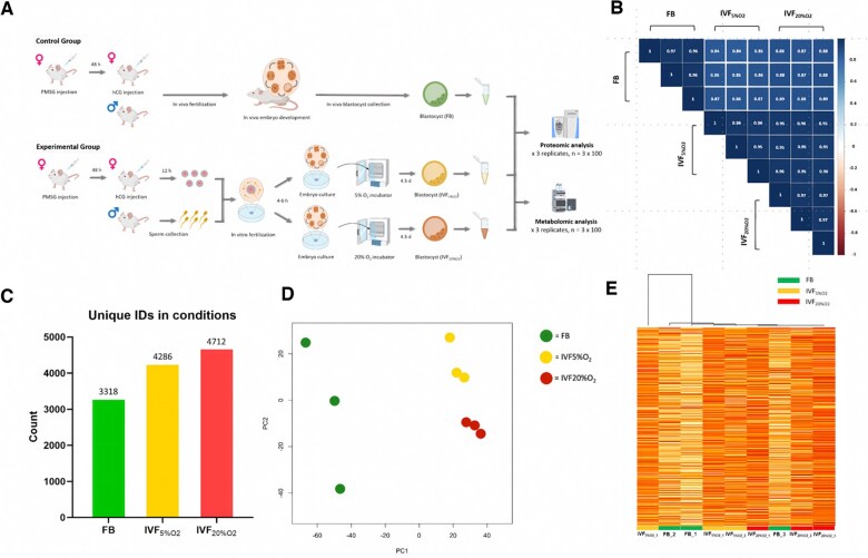
Study design size duration: Mouse embryos were generated by natural mating (in vivo control or flushed blastocyst-FB-group) or by IVF using KSOM medium and two distinct oxygen concentrations: 5% O2 (optimal) and 20% O2 (stressful). Proteomic and metabolomic analyses were performed using state-of-the-art mass spectrometry techniques in triplicate (n = 100 blastocysts per replicate), allowing for detailed profiling of protein and metabolite alterations in each group.
Participants/materials setting methods: Mouse blastocysts were collected from CD-1 and B6D2F1 strains as specified above. High-resolution liquid chromatography-tandem mass spectrometry (LC-MS/MS) was used for proteomics, while high-performance liquid chromatography coupled with mass spectrometry (HILIC-MS) was used for metabolomics. In addition, Immunofluorescence was used to assess the activation of stress response pathways, including the ISR.
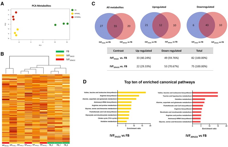
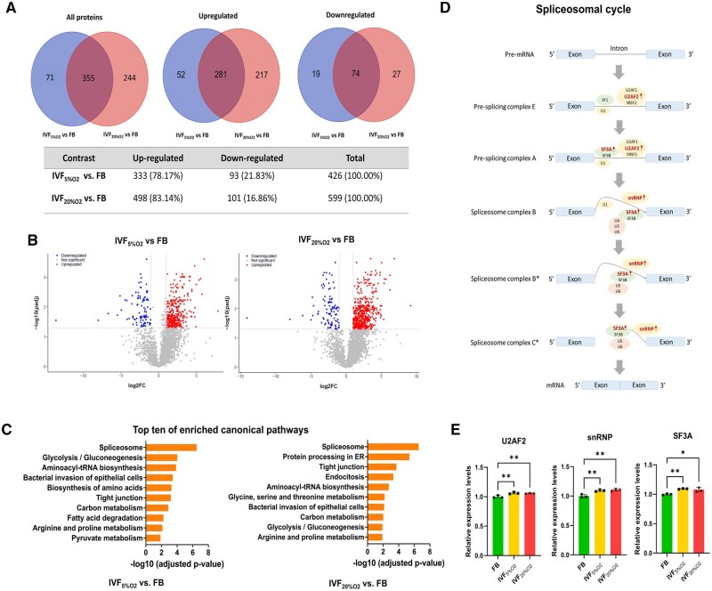
Main results and the role of chance: Proteomic analysis revealed significant changes in protein expression in embryos cultured under 20% O2 compared to 5% O2 and in vivo embryos. Compared to in vivo embryos, IVF embryos cultured under 20% O2 exhibited 599 differentially expressed proteins, with an increase in proteins involved in oxidative stress responses, aminoacyl-tRNA synthesis, and spliceosome pathways. In contrast, IVF embryos cultured under 5% O2 showed fewer changes, with 426 differentially expressed proteins, though still reflecting significant alterations compared to in vivo embryos. These results indicate that embryos in stressful conditions (20% O2) exhibit a stronger stress response and alterations in critical pathways for protein synthesis and DNA repair. Metabolomic analysis revealed that embryos cultured under 20% O2 showed changes in branch-chained amino acid levels, and decreased levels of key metabolites of the TCA cycle and pentose phosphate pathway. Embryos cultured under 5% O2 had increased pyruvate levels, suggesting altered glycolysis. Immunofluorescence confirmed that oxidative stress markers such as GCN2, EIF2α, and ATF4 were upregulated in IVF embryos, indicating ISR activation. Overall, IVF and embryo culture have a direct impact on embryo proteomes and metabolomes affecting amino acid metabolism and stress-related pathways.
Large scale data: N/A.
Limitations reasons for caution: Results in a murine model should be extrapolated with caution to human embryos.
Wider implications of the findings: These findings offer valuable insights into how different IVF culture conditions, specifically oxygen levels, impact the global metabolic and proteomic profiles of embryos. These findings provide critical insights into the profound impact of IVF culture conditions, particularly oxygen levels, on the global metabolic and proteomic landscapes of embryos. By identifying key metabolic pathways disrupted by oxidative stress, we highlight the potential clinical importance of proteomic and metabolomic analyses in understanding embryo quality, improving ART, and ultimately enhancing pregnancy outcomes. The integration of metabolomic and proteomic data offers a comprehensive understanding of how oxidative stress influences cellular function. These insights have direct clinical relevance, providing a foundation for optimizing ART protocols to mitigate oxidative stress.
Study funding/competing interests: This work was supported by grant R01 HD108166-01A1 from the National Institute of Child Health and Human Development (NICHD) to P.F.R. The authors declare that there is no conflict of interest that could be perceived as prejudicing the impartiality of the research reported.

 求助内容:
求助内容: 应助结果提醒方式:


