{"title":"肺腺癌固体成分的定性和定量CT分析预测侵袭性。","authors":"Yoshie Kunihiro, Fumi Kameda, Taiga Kobayashi, Masahiro Tanabe, Ryoko Morooka, Toshiki Tanaka, Yoshinobu Hoshii, Tsuneo Matsumoto, Katsuyoshi Ito","doi":"10.1007/s11604-025-01794-6","DOIUrl":null,"url":null,"abstract":"<p><strong>Purpose: </strong>This study aimed to evaluate the CT findings of lung adenocarcinoma with solid components and to determine the difference between adenocarcinoma in situ (AIS) and minimally invasive adenocarcinoma (MIA) with invasive adenocarcinoma (IAC).</p><p><strong>Materials and methods: </strong>A total of 54 cases were included in this study. The diagnoses of lung adenocarcinoma consisted of AIS or MIA (n = 20) and IAC (n = 34). The following factors were evaluated on CT images: part-solid nodule or solid nodule, presence of air bronchogram, air space, calcification within the tumor, presence of interstitial pneumonia and emphysema, diameters of the tumor and solid component, and CT values of the solid component. The volume and CT number histograms, including the 50th, 75th, and 100th percentiles of solid component were obtained using a software program. The CT criteria were compared between AIS, MIA, and IAC, and an indicator of differentiation was considered.</p><p><strong>Results: </strong>Part-solid nodules were observed more frequently in AIS and MIA (85.0%) than in IAC (55.9%). All criteria for quantitative analysis showed significant differences between AIS or MIA and IAC, and the diameter of the solid component in the mediastinal window was an indicator of differentiation (p = 0.0006; odds ratio, 1.4; 95% confidence interval, 1.2-1.8).</p><p><strong>Conclusion: </strong>The diameter of the solid component on the mediastinal window was considered an indicator of differentiation between AIS, MIA, and IAC.</p><p><strong>Condensed abstract: </strong>Quantitative data of solid component, including both manual measurements and evaluation using CT software, are correlated with pathological invasiveness. Diameter of the solid component in the mediastinal window would be an indicator of IAC.</p>","PeriodicalId":14691,"journal":{"name":"Japanese Journal of Radiology","volume":" ","pages":"1489-1497"},"PeriodicalIF":2.1000,"publicationDate":"2025-09-01","publicationTypes":"Journal Article","fieldsOfStudy":null,"isOpenAccess":false,"openAccessPdf":"https://www.ncbi.nlm.nih.gov/pmc/articles/PMC12397110/pdf/","citationCount":"0","resultStr":"{\"title\":\"Qualitative and quantitative CT analyses of the solid component in lung adenocarcinoma for predicting invasiveness.\",\"authors\":\"Yoshie Kunihiro, Fumi Kameda, Taiga Kobayashi, Masahiro Tanabe, Ryoko Morooka, Toshiki Tanaka, Yoshinobu Hoshii, Tsuneo Matsumoto, Katsuyoshi Ito\",\"doi\":\"10.1007/s11604-025-01794-6\",\"DOIUrl\":null,\"url\":null,\"abstract\":\"<p><strong>Purpose: </strong>This study aimed to evaluate the CT findings of lung adenocarcinoma with solid components and to determine the difference between adenocarcinoma in situ (AIS) and minimally invasive adenocarcinoma (MIA) with invasive adenocarcinoma (IAC).</p><p><strong>Materials and methods: </strong>A total of 54 cases were included in this study. The diagnoses of lung adenocarcinoma consisted of AIS or MIA (n = 20) and IAC (n = 34). The following factors were evaluated on CT images: part-solid nodule or solid nodule, presence of air bronchogram, air space, calcification within the tumor, presence of interstitial pneumonia and emphysema, diameters of the tumor and solid component, and CT values of the solid component. The volume and CT number histograms, including the 50th, 75th, and 100th percentiles of solid component were obtained using a software program. The CT criteria were compared between AIS, MIA, and IAC, and an indicator of differentiation was considered.</p><p><strong>Results: </strong>Part-solid nodules were observed more frequently in AIS and MIA (85.0%) than in IAC (55.9%). All criteria for quantitative analysis showed significant differences between AIS or MIA and IAC, and the diameter of the solid component in the mediastinal window was an indicator of differentiation (p = 0.0006; odds ratio, 1.4; 95% confidence interval, 1.2-1.8).</p><p><strong>Conclusion: </strong>The diameter of the solid component on the mediastinal window was considered an indicator of differentiation between AIS, MIA, and IAC.</p><p><strong>Condensed abstract: </strong>Quantitative data of solid component, including both manual measurements and evaluation using CT software, are correlated with pathological invasiveness. Diameter of the solid component in the mediastinal window would be an indicator of IAC.</p>\",\"PeriodicalId\":14691,\"journal\":{\"name\":\"Japanese Journal of Radiology\",\"volume\":\" \",\"pages\":\"1489-1497\"},\"PeriodicalIF\":2.1000,\"publicationDate\":\"2025-09-01\",\"publicationTypes\":\"Journal Article\",\"fieldsOfStudy\":null,\"isOpenAccess\":false,\"openAccessPdf\":\"https://www.ncbi.nlm.nih.gov/pmc/articles/PMC12397110/pdf/\",\"citationCount\":\"0\",\"resultStr\":null,\"platform\":\"Semanticscholar\",\"paperid\":null,\"PeriodicalName\":\"Japanese Journal of Radiology\",\"FirstCategoryId\":\"3\",\"ListUrlMain\":\"https://doi.org/10.1007/s11604-025-01794-6\",\"RegionNum\":4,\"RegionCategory\":\"医学\",\"ArticlePicture\":[],\"TitleCN\":null,\"AbstractTextCN\":null,\"PMCID\":null,\"EPubDate\":\"2025/5/25 0:00:00\",\"PubModel\":\"Epub\",\"JCR\":\"\",\"JCRName\":\"\",\"Score\":null,\"Total\":0}","platform":"Semanticscholar","paperid":null,"PeriodicalName":"Japanese Journal of Radiology","FirstCategoryId":"3","ListUrlMain":"https://doi.org/10.1007/s11604-025-01794-6","RegionNum":4,"RegionCategory":"医学","ArticlePicture":[],"TitleCN":null,"AbstractTextCN":null,"PMCID":null,"EPubDate":"2025/5/25 0:00:00","PubModel":"Epub","JCR":"","JCRName":"","Score":null,"Total":0}
Qualitative and quantitative CT analyses of the solid component in lung adenocarcinoma for predicting invasiveness.
Purpose: This study aimed to evaluate the CT findings of lung adenocarcinoma with solid components and to determine the difference between adenocarcinoma in situ (AIS) and minimally invasive adenocarcinoma (MIA) with invasive adenocarcinoma (IAC).
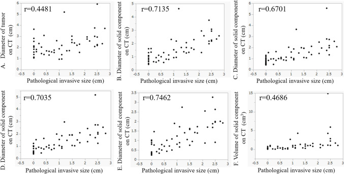
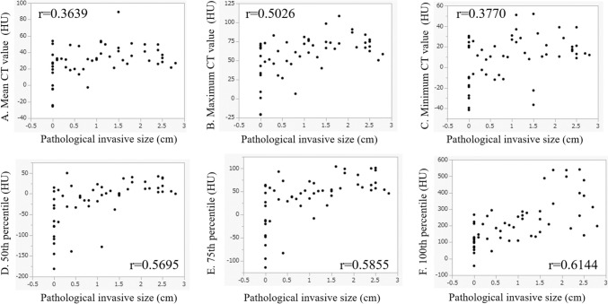
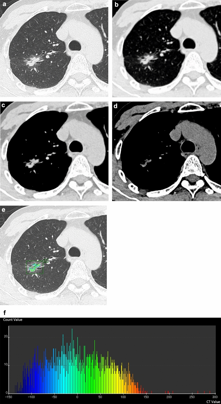
Materials and methods: A total of 54 cases were included in this study. The diagnoses of lung adenocarcinoma consisted of AIS or MIA (n = 20) and IAC (n = 34). The following factors were evaluated on CT images: part-solid nodule or solid nodule, presence of air bronchogram, air space, calcification within the tumor, presence of interstitial pneumonia and emphysema, diameters of the tumor and solid component, and CT values of the solid component. The volume and CT number histograms, including the 50th, 75th, and 100th percentiles of solid component were obtained using a software program. The CT criteria were compared between AIS, MIA, and IAC, and an indicator of differentiation was considered.
Results: Part-solid nodules were observed more frequently in AIS and MIA (85.0%) than in IAC (55.9%). All criteria for quantitative analysis showed significant differences between AIS or MIA and IAC, and the diameter of the solid component in the mediastinal window was an indicator of differentiation (p = 0.0006; odds ratio, 1.4; 95% confidence interval, 1.2-1.8).
Conclusion: The diameter of the solid component on the mediastinal window was considered an indicator of differentiation between AIS, MIA, and IAC.
Condensed abstract: Quantitative data of solid component, including both manual measurements and evaluation using CT software, are correlated with pathological invasiveness. Diameter of the solid component in the mediastinal window would be an indicator of IAC.
期刊介绍:
Japanese Journal of Radiology is a peer-reviewed journal, officially published by the Japan Radiological Society. The main purpose of the journal is to provide a forum for the publication of papers documenting recent advances and new developments in the field of radiology in medicine and biology. The scope of Japanese Journal of Radiology encompasses but is not restricted to diagnostic radiology, interventional radiology, radiation oncology, nuclear medicine, radiation physics, and radiation biology. Additionally, the journal covers technical and industrial innovations. The journal welcomes original articles, technical notes, review articles, pictorial essays and letters to the editor. The journal also provides announcements from the boards and the committees of the society. Membership in the Japan Radiological Society is not a prerequisite for submission. Contributions are welcomed from all parts of the world.

 求助内容:
求助内容: 应助结果提醒方式:


