Maya Abi-Akl, Jens Maebe, Boris Vervenne, Othmane Bouhali, Christian Vanhove, Stefaan Vandenberghe
{"title":"基于单片LYSO探测器平板的中轴视场稀疏PET系统性能评价:仿真研究。","authors":"Maya Abi-Akl, Jens Maebe, Boris Vervenne, Othmane Bouhali, Christian Vanhove, Stefaan Vandenberghe","doi":"10.1186/s40658-025-00766-z","DOIUrl":null,"url":null,"abstract":"<p><strong>Background: </strong>The combination of longer axial field-of-view (AFOV) and time-of-flight positron emission tomography (PET) has significantly improved system sensitivity and, as a result, image quality. This study investigates a cost-effective extended AFOV PET system design using monolithic LYSO detectors with depth-of-interaction capabilities. These detectors, arranged in a vertical flat-panel geometry and positioned closer to the patient, enable superior spatial resolution while maintaining a compact and affordable system design. We simulate the performance of two flat-panel PET configurations: one with a fully populated 106 cm AFOV and another cost-efficient design featuring a reduced AFOV with axial gaps and vertical panel motion optimized for head and torso imaging.</p><p><strong>Methods: </strong>Both configurations consist of two monolithic LYSO-based flat panels placed 50 cm apart. The panels are 71 cm wide, with the Long Flat Panel (L-FP) design extending to a length of 106 cm while the Sparse Medium Flat Panel (SpM-FP) design measures 60 cm in length. Monte Carlo simulations evaluated the two designs using the NEMA protocol and additional tests for a more thorough assessment. Sensitivity, spatial resolution, axial noise variability, and image quality were analyzed, and an XCAT phantom at standard dose was used to demonstrate the achievable clinical image quality.</p><p><strong>Results: </strong>The SpM-FP showed 4-5 times lower sensitivity than the L-FP, requiring an acquisition time of 2-3 min to match the image quality achieved by the L-FP in 30 s. This finding is supported by the contrast-to-noise ratio of the image quality phantom and the standard deviation values obtained from the liver and lung regions of the XCAT phantom. Both configurations achieved uniform spatial resolution below 2 mm in the two directions parallel to the panels and an average of 3-3.5 mm in the direction towards the panels, with slight degradation observed away from the center of the AFOV. Additionally, the axial noise profile of the SpM-FP revealed minimal variability.</p><p><strong>Conclusions: </strong>The SpM-FP design shows potential as a cost-effective system, combining the benefits of extended AFOV, superior spatial resolution and high patient throughput.</p>","PeriodicalId":11559,"journal":{"name":"EJNMMI Physics","volume":"12 1","pages":"49"},"PeriodicalIF":3.2000,"publicationDate":"2025-05-26","publicationTypes":"Journal Article","fieldsOfStudy":null,"isOpenAccess":false,"openAccessPdf":"https://www.ncbi.nlm.nih.gov/pmc/articles/PMC12106179/pdf/","citationCount":"0","resultStr":"{\"title\":\"Performance evaluation of a medium axial field-of-view sparse PET system based on flat panels of monolithic LYSO detectors: a simulation study.\",\"authors\":\"Maya Abi-Akl, Jens Maebe, Boris Vervenne, Othmane Bouhali, Christian Vanhove, Stefaan Vandenberghe\",\"doi\":\"10.1186/s40658-025-00766-z\",\"DOIUrl\":null,\"url\":null,\"abstract\":\"<p><strong>Background: </strong>The combination of longer axial field-of-view (AFOV) and time-of-flight positron emission tomography (PET) has significantly improved system sensitivity and, as a result, image quality. This study investigates a cost-effective extended AFOV PET system design using monolithic LYSO detectors with depth-of-interaction capabilities. These detectors, arranged in a vertical flat-panel geometry and positioned closer to the patient, enable superior spatial resolution while maintaining a compact and affordable system design. We simulate the performance of two flat-panel PET configurations: one with a fully populated 106 cm AFOV and another cost-efficient design featuring a reduced AFOV with axial gaps and vertical panel motion optimized for head and torso imaging.</p><p><strong>Methods: </strong>Both configurations consist of two monolithic LYSO-based flat panels placed 50 cm apart. The panels are 71 cm wide, with the Long Flat Panel (L-FP) design extending to a length of 106 cm while the Sparse Medium Flat Panel (SpM-FP) design measures 60 cm in length. Monte Carlo simulations evaluated the two designs using the NEMA protocol and additional tests for a more thorough assessment. Sensitivity, spatial resolution, axial noise variability, and image quality were analyzed, and an XCAT phantom at standard dose was used to demonstrate the achievable clinical image quality.</p><p><strong>Results: </strong>The SpM-FP showed 4-5 times lower sensitivity than the L-FP, requiring an acquisition time of 2-3 min to match the image quality achieved by the L-FP in 30 s. This finding is supported by the contrast-to-noise ratio of the image quality phantom and the standard deviation values obtained from the liver and lung regions of the XCAT phantom. Both configurations achieved uniform spatial resolution below 2 mm in the two directions parallel to the panels and an average of 3-3.5 mm in the direction towards the panels, with slight degradation observed away from the center of the AFOV. Additionally, the axial noise profile of the SpM-FP revealed minimal variability.</p><p><strong>Conclusions: </strong>The SpM-FP design shows potential as a cost-effective system, combining the benefits of extended AFOV, superior spatial resolution and high patient throughput.</p>\",\"PeriodicalId\":11559,\"journal\":{\"name\":\"EJNMMI Physics\",\"volume\":\"12 1\",\"pages\":\"49\"},\"PeriodicalIF\":3.2000,\"publicationDate\":\"2025-05-26\",\"publicationTypes\":\"Journal Article\",\"fieldsOfStudy\":null,\"isOpenAccess\":false,\"openAccessPdf\":\"https://www.ncbi.nlm.nih.gov/pmc/articles/PMC12106179/pdf/\",\"citationCount\":\"0\",\"resultStr\":null,\"platform\":\"Semanticscholar\",\"paperid\":null,\"PeriodicalName\":\"EJNMMI Physics\",\"FirstCategoryId\":\"3\",\"ListUrlMain\":\"https://doi.org/10.1186/s40658-025-00766-z\",\"RegionNum\":2,\"RegionCategory\":\"医学\",\"ArticlePicture\":[],\"TitleCN\":null,\"AbstractTextCN\":null,\"PMCID\":null,\"EPubDate\":\"\",\"PubModel\":\"\",\"JCR\":\"Q2\",\"JCRName\":\"RADIOLOGY, NUCLEAR MEDICINE & MEDICAL IMAGING\",\"Score\":null,\"Total\":0}","platform":"Semanticscholar","paperid":null,"PeriodicalName":"EJNMMI Physics","FirstCategoryId":"3","ListUrlMain":"https://doi.org/10.1186/s40658-025-00766-z","RegionNum":2,"RegionCategory":"医学","ArticlePicture":[],"TitleCN":null,"AbstractTextCN":null,"PMCID":null,"EPubDate":"","PubModel":"","JCR":"Q2","JCRName":"RADIOLOGY, NUCLEAR MEDICINE & MEDICAL IMAGING","Score":null,"Total":0}
Performance evaluation of a medium axial field-of-view sparse PET system based on flat panels of monolithic LYSO detectors: a simulation study.
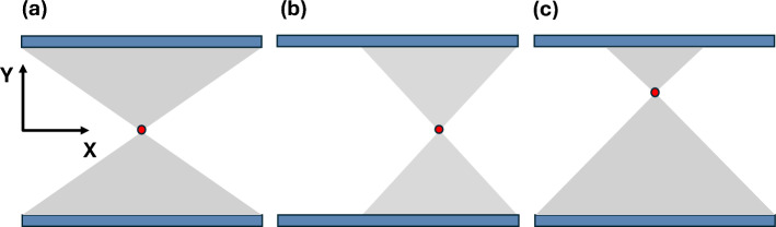
Background: The combination of longer axial field-of-view (AFOV) and time-of-flight positron emission tomography (PET) has significantly improved system sensitivity and, as a result, image quality. This study investigates a cost-effective extended AFOV PET system design using monolithic LYSO detectors with depth-of-interaction capabilities. These detectors, arranged in a vertical flat-panel geometry and positioned closer to the patient, enable superior spatial resolution while maintaining a compact and affordable system design. We simulate the performance of two flat-panel PET configurations: one with a fully populated 106 cm AFOV and another cost-efficient design featuring a reduced AFOV with axial gaps and vertical panel motion optimized for head and torso imaging.
Methods: Both configurations consist of two monolithic LYSO-based flat panels placed 50 cm apart. The panels are 71 cm wide, with the Long Flat Panel (L-FP) design extending to a length of 106 cm while the Sparse Medium Flat Panel (SpM-FP) design measures 60 cm in length. Monte Carlo simulations evaluated the two designs using the NEMA protocol and additional tests for a more thorough assessment. Sensitivity, spatial resolution, axial noise variability, and image quality were analyzed, and an XCAT phantom at standard dose was used to demonstrate the achievable clinical image quality.
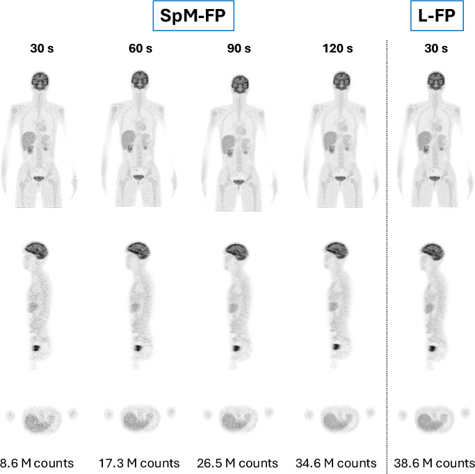
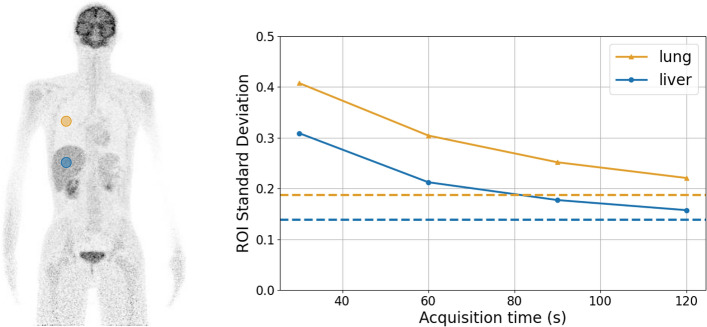
Results: The SpM-FP showed 4-5 times lower sensitivity than the L-FP, requiring an acquisition time of 2-3 min to match the image quality achieved by the L-FP in 30 s. This finding is supported by the contrast-to-noise ratio of the image quality phantom and the standard deviation values obtained from the liver and lung regions of the XCAT phantom. Both configurations achieved uniform spatial resolution below 2 mm in the two directions parallel to the panels and an average of 3-3.5 mm in the direction towards the panels, with slight degradation observed away from the center of the AFOV. Additionally, the axial noise profile of the SpM-FP revealed minimal variability.
Conclusions: The SpM-FP design shows potential as a cost-effective system, combining the benefits of extended AFOV, superior spatial resolution and high patient throughput.
期刊介绍:
EJNMMI Physics is an international platform for scientists, users and adopters of nuclear medicine with a particular interest in physics matters. As a companion journal to the European Journal of Nuclear Medicine and Molecular Imaging, this journal has a multi-disciplinary approach and welcomes original materials and studies with a focus on applied physics and mathematics as well as imaging systems engineering and prototyping in nuclear medicine. This includes physics-driven approaches or algorithms supported by physics that foster early clinical adoption of nuclear medicine imaging and therapy.

 求助内容:
求助内容: 应助结果提醒方式:


