Nikolaos Kathopoulis, Konstantinos Kypriotis, Athanasios Douligeris, Michael Panagiotopouloz, Ioannis Chatzipapas, Athanasios Protopapas
{"title":"剖宫产瘢痕妊娠的腹腔镜治疗10步。","authors":"Nikolaos Kathopoulis, Konstantinos Kypriotis, Athanasios Douligeris, Michael Panagiotopouloz, Ioannis Chatzipapas, Athanasios Protopapas","doi":"10.52054/FVVO.2025.14","DOIUrl":null,"url":null,"abstract":"<p><strong>Background: </strong>Caesarean scar pregnancy (CSP) is a pathologic entity with rising incidence over recent years. So far, there are many treatment methods and protocols suggesting surgical or medical interventions and their combinations. More and more laparoscopic surgery is applied to treat scar pregnancy with excellent results. A proper surgical strategy is a key point for optimal surgical outcome.</p><p><strong>Objectives: </strong>To present a standardised technique for the laparoscopic management of CSP.</p><p><strong>Participant: </strong>Patients with CSP having the indication of laparoscopic treatment.</p><p><strong>Intervention: </strong>The video presents a systematic approach of the laparoscopic treatment of CSP clearly divided into 10 steps: 1. Prepare the surgery; 2. Inspection of the pelvis; 3. Bladder dissection; 4. Preventive haemostasis; 5. Hysterotomy; 6. Evacuation of conception products; 7. Excision of niche scar tissue; 8. Evacuation of the uterine cavity; 9. Suturing of the uterine defect; 10. Removal of the uterine artery clips. The main outcome measures are the efficacy of the laparoscopic management of CSP and the postoperative uterine reconstruction in terms of ultrasonic measurement of the isthmic myometrial layer thickness. Patients are released from the hospital the day after the surgery, and a follow-up ultrasound is scheduled three months post-operatively. In the case presented in the video, the myometrial wall is increased from 3 mm preoperatively to 13 mm three months postoperatively.</p><p><strong>Conclusions: </strong>The main advantage of this technique is the ability to treat CSP, remove the uterine isthmocele, and reconstruct the lower uterine segment simultaneously. The 10 steps proposed in a logical sequence may shorten the surgery's learning curve and reduce possible complications.</p><p><strong>What is new?: </strong>We present a systematic approach that provides a safe and easily reproducible technique for managing CSP.</p>","PeriodicalId":46400,"journal":{"name":"Facts Views and Vision in ObGyn","volume":" ","pages":"208-211"},"PeriodicalIF":1.4000,"publicationDate":"2025-06-27","publicationTypes":"Journal Article","fieldsOfStudy":null,"isOpenAccess":false,"openAccessPdf":"https://www.ncbi.nlm.nih.gov/pmc/articles/PMC12233120/pdf/","citationCount":"0","resultStr":"{\"title\":\"Laparoscopic management of caesarean scar pregnancy in 10 steps.\",\"authors\":\"Nikolaos Kathopoulis, Konstantinos Kypriotis, Athanasios Douligeris, Michael Panagiotopouloz, Ioannis Chatzipapas, Athanasios Protopapas\",\"doi\":\"10.52054/FVVO.2025.14\",\"DOIUrl\":null,\"url\":null,\"abstract\":\"<p><strong>Background: </strong>Caesarean scar pregnancy (CSP) is a pathologic entity with rising incidence over recent years. So far, there are many treatment methods and protocols suggesting surgical or medical interventions and their combinations. More and more laparoscopic surgery is applied to treat scar pregnancy with excellent results. A proper surgical strategy is a key point for optimal surgical outcome.</p><p><strong>Objectives: </strong>To present a standardised technique for the laparoscopic management of CSP.</p><p><strong>Participant: </strong>Patients with CSP having the indication of laparoscopic treatment.</p><p><strong>Intervention: </strong>The video presents a systematic approach of the laparoscopic treatment of CSP clearly divided into 10 steps: 1. Prepare the surgery; 2. Inspection of the pelvis; 3. Bladder dissection; 4. Preventive haemostasis; 5. Hysterotomy; 6. Evacuation of conception products; 7. Excision of niche scar tissue; 8. Evacuation of the uterine cavity; 9. Suturing of the uterine defect; 10. Removal of the uterine artery clips. The main outcome measures are the efficacy of the laparoscopic management of CSP and the postoperative uterine reconstruction in terms of ultrasonic measurement of the isthmic myometrial layer thickness. Patients are released from the hospital the day after the surgery, and a follow-up ultrasound is scheduled three months post-operatively. In the case presented in the video, the myometrial wall is increased from 3 mm preoperatively to 13 mm three months postoperatively.</p><p><strong>Conclusions: </strong>The main advantage of this technique is the ability to treat CSP, remove the uterine isthmocele, and reconstruct the lower uterine segment simultaneously. The 10 steps proposed in a logical sequence may shorten the surgery's learning curve and reduce possible complications.</p><p><strong>What is new?: </strong>We present a systematic approach that provides a safe and easily reproducible technique for managing CSP.</p>\",\"PeriodicalId\":46400,\"journal\":{\"name\":\"Facts Views and Vision in ObGyn\",\"volume\":\" \",\"pages\":\"208-211\"},\"PeriodicalIF\":1.4000,\"publicationDate\":\"2025-06-27\",\"publicationTypes\":\"Journal Article\",\"fieldsOfStudy\":null,\"isOpenAccess\":false,\"openAccessPdf\":\"https://www.ncbi.nlm.nih.gov/pmc/articles/PMC12233120/pdf/\",\"citationCount\":\"0\",\"resultStr\":null,\"platform\":\"Semanticscholar\",\"paperid\":null,\"PeriodicalName\":\"Facts Views and Vision in ObGyn\",\"FirstCategoryId\":\"1085\",\"ListUrlMain\":\"https://doi.org/10.52054/FVVO.2025.14\",\"RegionNum\":0,\"RegionCategory\":null,\"ArticlePicture\":[],\"TitleCN\":null,\"AbstractTextCN\":null,\"PMCID\":null,\"EPubDate\":\"2025/5/23 0:00:00\",\"PubModel\":\"Epub\",\"JCR\":\"Q3\",\"JCRName\":\"OBSTETRICS & GYNECOLOGY\",\"Score\":null,\"Total\":0}","platform":"Semanticscholar","paperid":null,"PeriodicalName":"Facts Views and Vision in ObGyn","FirstCategoryId":"1085","ListUrlMain":"https://doi.org/10.52054/FVVO.2025.14","RegionNum":0,"RegionCategory":null,"ArticlePicture":[],"TitleCN":null,"AbstractTextCN":null,"PMCID":null,"EPubDate":"2025/5/23 0:00:00","PubModel":"Epub","JCR":"Q3","JCRName":"OBSTETRICS & GYNECOLOGY","Score":null,"Total":0}
Laparoscopic management of caesarean scar pregnancy in 10 steps.
Background: Caesarean scar pregnancy (CSP) is a pathologic entity with rising incidence over recent years. So far, there are many treatment methods and protocols suggesting surgical or medical interventions and their combinations. More and more laparoscopic surgery is applied to treat scar pregnancy with excellent results. A proper surgical strategy is a key point for optimal surgical outcome.
Objectives: To present a standardised technique for the laparoscopic management of CSP.
Participant: Patients with CSP having the indication of laparoscopic treatment.
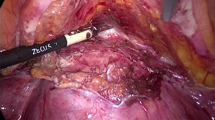
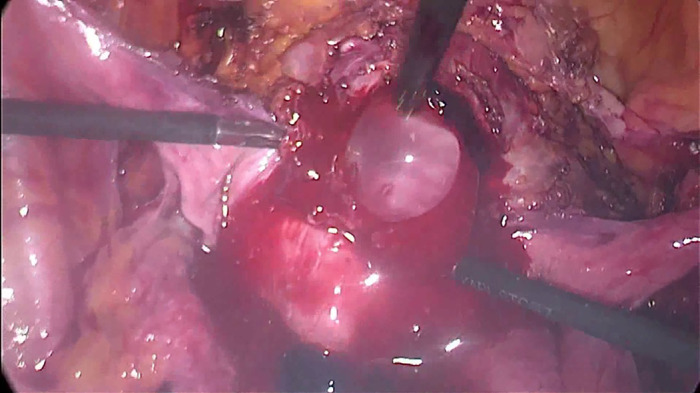
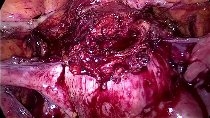
Intervention: The video presents a systematic approach of the laparoscopic treatment of CSP clearly divided into 10 steps: 1. Prepare the surgery; 2. Inspection of the pelvis; 3. Bladder dissection; 4. Preventive haemostasis; 5. Hysterotomy; 6. Evacuation of conception products; 7. Excision of niche scar tissue; 8. Evacuation of the uterine cavity; 9. Suturing of the uterine defect; 10. Removal of the uterine artery clips. The main outcome measures are the efficacy of the laparoscopic management of CSP and the postoperative uterine reconstruction in terms of ultrasonic measurement of the isthmic myometrial layer thickness. Patients are released from the hospital the day after the surgery, and a follow-up ultrasound is scheduled three months post-operatively. In the case presented in the video, the myometrial wall is increased from 3 mm preoperatively to 13 mm three months postoperatively.
Conclusions: The main advantage of this technique is the ability to treat CSP, remove the uterine isthmocele, and reconstruct the lower uterine segment simultaneously. The 10 steps proposed in a logical sequence may shorten the surgery's learning curve and reduce possible complications.
What is new?: We present a systematic approach that provides a safe and easily reproducible technique for managing CSP.

 求助内容:
求助内容: 应助结果提醒方式:


