François Cousin, Thomas Louis, Pierre Frères, Julien Guiot, Mariaelena Occhipinti, Fabio Bottari, Wim Vos, Roland Hustinx
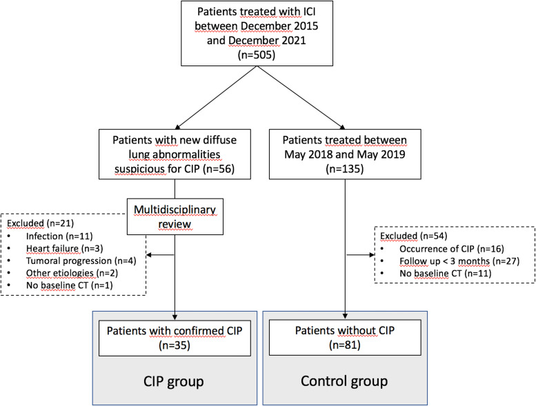
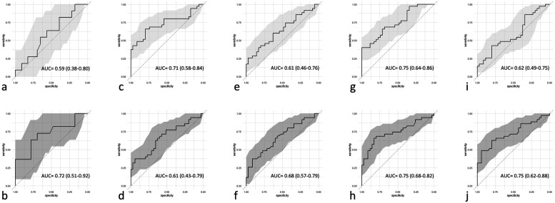
{"title":"整合肺部CT放射组学的机器学习模型预测晚期癌症患者的检查点抑制剂肺炎。","authors":"François Cousin, Thomas Louis, Pierre Frères, Julien Guiot, Mariaelena Occhipinti, Fabio Bottari, Wim Vos, Roland Hustinx","doi":"10.1177/15330338251344004","DOIUrl":null,"url":null,"abstract":"<p><p>ObjectiveCheckpoint inhibitor pneumonitis (CIP) is a potentially life-threatening immune-related adverse event. Efficient strategies to select patients at risk are still required. The aim of our study was to assess the utility of a machine learning model, integrating pre-treatment CT lung radiomics features with clinical data, to predict patients at risk of developing CIP.MethodsIn this retrospective study, 116 patients with varied malignancies treated with immune checkpoint inhibitors (ICIs) were included. In this cohort, 35 patients presented with CIP and 81 patients did not. Each lung and its lobes were segmented on pre-treatment CT scans to perform a handcrafted radiomic analysis. Radiomic features were associated with clinical parameters to build generalized linear (GLM) and random forest (RF) models, to predict occurrence of CIP. The models were fine-tuned, validated and tested using a nested 5-fold cross-validation method.ResultsThe RF models combining radiomic and clinical features showed the best performances with an area under the ROC curve (AUC) of 0.75 (95%CI:0.62-0.88) on the test set. The most accurate clinical model was a RF model and achieved an AUC of 0.72 (95%CI:0.51-0.92). The best radiomic model was a GLM model and achieved an AUC of 0.71 (95%CI:0.58-0.84).ConclusionsOur CT-based lung radiomic models showed moderate to good performance at predicting CIP. We demonstrated the potential role of machine learning models associating clinical parameters and lung CT radiomic features to better identify patients treated with ICIs at risk of developing CIP.Advances in knowledge: Radiomics analysis of the lung parenchyma could be used as a non-invasive tool to select patients at risk of developing immune-checkpoint pneumonitis.</p>","PeriodicalId":22203,"journal":{"name":"Technology in Cancer Research & Treatment","volume":"24 ","pages":"15330338251344004"},"PeriodicalIF":2.8000,"publicationDate":"2025-01-01","publicationTypes":"Journal Article","fieldsOfStudy":null,"isOpenAccess":false,"openAccessPdf":"https://www.ncbi.nlm.nih.gov/pmc/articles/PMC12102562/pdf/","citationCount":"0","resultStr":"{\"title\":\"Machine Learning Model Integrating CT Radiomics of the Lung to Predict Checkpoint Inhibitor Pneumonitis in Patients with Advanced Cancer.\",\"authors\":\"François Cousin, Thomas Louis, Pierre Frères, Julien Guiot, Mariaelena Occhipinti, Fabio Bottari, Wim Vos, Roland Hustinx\",\"doi\":\"10.1177/15330338251344004\",\"DOIUrl\":null,\"url\":null,\"abstract\":\"<p><p>ObjectiveCheckpoint inhibitor pneumonitis (CIP) is a potentially life-threatening immune-related adverse event. Efficient strategies to select patients at risk are still required. The aim of our study was to assess the utility of a machine learning model, integrating pre-treatment CT lung radiomics features with clinical data, to predict patients at risk of developing CIP.MethodsIn this retrospective study, 116 patients with varied malignancies treated with immune checkpoint inhibitors (ICIs) were included. In this cohort, 35 patients presented with CIP and 81 patients did not. Each lung and its lobes were segmented on pre-treatment CT scans to perform a handcrafted radiomic analysis. Radiomic features were associated with clinical parameters to build generalized linear (GLM) and random forest (RF) models, to predict occurrence of CIP. The models were fine-tuned, validated and tested using a nested 5-fold cross-validation method.ResultsThe RF models combining radiomic and clinical features showed the best performances with an area under the ROC curve (AUC) of 0.75 (95%CI:0.62-0.88) on the test set. The most accurate clinical model was a RF model and achieved an AUC of 0.72 (95%CI:0.51-0.92). The best radiomic model was a GLM model and achieved an AUC of 0.71 (95%CI:0.58-0.84).ConclusionsOur CT-based lung radiomic models showed moderate to good performance at predicting CIP. We demonstrated the potential role of machine learning models associating clinical parameters and lung CT radiomic features to better identify patients treated with ICIs at risk of developing CIP.Advances in knowledge: Radiomics analysis of the lung parenchyma could be used as a non-invasive tool to select patients at risk of developing immune-checkpoint pneumonitis.</p>\",\"PeriodicalId\":22203,\"journal\":{\"name\":\"Technology in Cancer Research & Treatment\",\"volume\":\"24 \",\"pages\":\"15330338251344004\"},\"PeriodicalIF\":2.8000,\"publicationDate\":\"2025-01-01\",\"publicationTypes\":\"Journal Article\",\"fieldsOfStudy\":null,\"isOpenAccess\":false,\"openAccessPdf\":\"https://www.ncbi.nlm.nih.gov/pmc/articles/PMC12102562/pdf/\",\"citationCount\":\"0\",\"resultStr\":null,\"platform\":\"Semanticscholar\",\"paperid\":null,\"PeriodicalName\":\"Technology in Cancer Research & Treatment\",\"FirstCategoryId\":\"3\",\"ListUrlMain\":\"https://doi.org/10.1177/15330338251344004\",\"RegionNum\":4,\"RegionCategory\":\"医学\",\"ArticlePicture\":[],\"TitleCN\":null,\"AbstractTextCN\":null,\"PMCID\":null,\"EPubDate\":\"2025/5/23 0:00:00\",\"PubModel\":\"Epub\",\"JCR\":\"Q3\",\"JCRName\":\"ONCOLOGY\",\"Score\":null,\"Total\":0}","platform":"Semanticscholar","paperid":null,"PeriodicalName":"Technology in Cancer Research & Treatment","FirstCategoryId":"3","ListUrlMain":"https://doi.org/10.1177/15330338251344004","RegionNum":4,"RegionCategory":"医学","ArticlePicture":[],"TitleCN":null,"AbstractTextCN":null,"PMCID":null,"EPubDate":"2025/5/23 0:00:00","PubModel":"Epub","JCR":"Q3","JCRName":"ONCOLOGY","Score":null,"Total":0}
Machine Learning Model Integrating CT Radiomics of the Lung to Predict Checkpoint Inhibitor Pneumonitis in Patients with Advanced Cancer.
ObjectiveCheckpoint inhibitor pneumonitis (CIP) is a potentially life-threatening immune-related adverse event. Efficient strategies to select patients at risk are still required. The aim of our study was to assess the utility of a machine learning model, integrating pre-treatment CT lung radiomics features with clinical data, to predict patients at risk of developing CIP.MethodsIn this retrospective study, 116 patients with varied malignancies treated with immune checkpoint inhibitors (ICIs) were included. In this cohort, 35 patients presented with CIP and 81 patients did not. Each lung and its lobes were segmented on pre-treatment CT scans to perform a handcrafted radiomic analysis. Radiomic features were associated with clinical parameters to build generalized linear (GLM) and random forest (RF) models, to predict occurrence of CIP. The models were fine-tuned, validated and tested using a nested 5-fold cross-validation method.ResultsThe RF models combining radiomic and clinical features showed the best performances with an area under the ROC curve (AUC) of 0.75 (95%CI:0.62-0.88) on the test set. The most accurate clinical model was a RF model and achieved an AUC of 0.72 (95%CI:0.51-0.92). The best radiomic model was a GLM model and achieved an AUC of 0.71 (95%CI:0.58-0.84).ConclusionsOur CT-based lung radiomic models showed moderate to good performance at predicting CIP. We demonstrated the potential role of machine learning models associating clinical parameters and lung CT radiomic features to better identify patients treated with ICIs at risk of developing CIP.Advances in knowledge: Radiomics analysis of the lung parenchyma could be used as a non-invasive tool to select patients at risk of developing immune-checkpoint pneumonitis.
期刊介绍:
Technology in Cancer Research & Treatment (TCRT) is a JCR-ranked, broad-spectrum, open access, peer-reviewed publication whose aim is to provide researchers and clinicians with a platform to share and discuss developments in the prevention, diagnosis, treatment, and monitoring of cancer.

 求助内容:
求助内容: 应助结果提醒方式:


