Mohammed Saleh, Islam Jadallah, Qais Alhroub, Maaweya Jabareen, Wasef Alhroub, Ammar Hassouneh
{"title":"微创隔肌切除术后肺疝:一例成功手术修复的罕见病例报告。","authors":"Mohammed Saleh, Islam Jadallah, Qais Alhroub, Maaweya Jabareen, Wasef Alhroub, Ammar Hassouneh","doi":"10.1016/j.ijscr.2025.111313","DOIUrl":null,"url":null,"abstract":"<p><strong>Background: </strong>Lung herniation is a rare condition where lung tissue protrudes through the thoracic wall, often due to surgery, trauma, or pressure changes. The intercostal type is most common, especially after minimally invasive cardiac surgery (MICS) like septal myectomy for HOCM. Early diagnosis and treatment are crucial to prevent complications like respiratory distress and lung strangulation.</p><p><strong>Case presentation: </strong>A 57-year-old male with cardiac disease underwent minimally invasive septal myectomy for HOCM. Ten days later, he developed shortness of breath, dry cough, and a right chest mass. Imaging studies, including a chest X-ray revealed a triangular lucency lateral to the right 4th and 5th ribs and non-contrast CT confirmed lung herniation due to an intercostal muscle tear. He underwent surgical repair with rib plating and mesh, leading to significant improvement. He was discharged after seven days and remained asymptomatic at 8-month follow-up.</p><p><strong>Discussion: </strong>Lung herniation after MICS is rare but serious, caused by intercostal muscle injury. Risk factors include lung conditions, pressure changes, and surgery. Small cases may resolve, but larger ones need surgery. Timely imaging and rib plating with mesh prevented complications and ensured recovery.</p><p><strong>Conclusion: </strong>Lung herniation should be considered in post-MICS patients with respiratory distress and chest abnormalities. Early diagnosis and surgery are crucial to prevent complications. This case emphasizes the importance of prompt recognition and proper surgical management.</p>","PeriodicalId":48113,"journal":{"name":"International Journal of Surgery Case Reports","volume":"131 ","pages":"111313"},"PeriodicalIF":0.7000,"publicationDate":"2025-06-01","publicationTypes":"Journal Article","fieldsOfStudy":null,"isOpenAccess":false,"openAccessPdf":"https://www.ncbi.nlm.nih.gov/pmc/articles/PMC12145991/pdf/","citationCount":"0","resultStr":"{\"title\":\"Postoperative lung herniation following minimally invasive septal myectomy: A rare case report with successful surgical repair.\",\"authors\":\"Mohammed Saleh, Islam Jadallah, Qais Alhroub, Maaweya Jabareen, Wasef Alhroub, Ammar Hassouneh\",\"doi\":\"10.1016/j.ijscr.2025.111313\",\"DOIUrl\":null,\"url\":null,\"abstract\":\"<p><strong>Background: </strong>Lung herniation is a rare condition where lung tissue protrudes through the thoracic wall, often due to surgery, trauma, or pressure changes. The intercostal type is most common, especially after minimally invasive cardiac surgery (MICS) like septal myectomy for HOCM. Early diagnosis and treatment are crucial to prevent complications like respiratory distress and lung strangulation.</p><p><strong>Case presentation: </strong>A 57-year-old male with cardiac disease underwent minimally invasive septal myectomy for HOCM. Ten days later, he developed shortness of breath, dry cough, and a right chest mass. Imaging studies, including a chest X-ray revealed a triangular lucency lateral to the right 4th and 5th ribs and non-contrast CT confirmed lung herniation due to an intercostal muscle tear. He underwent surgical repair with rib plating and mesh, leading to significant improvement. He was discharged after seven days and remained asymptomatic at 8-month follow-up.</p><p><strong>Discussion: </strong>Lung herniation after MICS is rare but serious, caused by intercostal muscle injury. Risk factors include lung conditions, pressure changes, and surgery. Small cases may resolve, but larger ones need surgery. Timely imaging and rib plating with mesh prevented complications and ensured recovery.</p><p><strong>Conclusion: </strong>Lung herniation should be considered in post-MICS patients with respiratory distress and chest abnormalities. Early diagnosis and surgery are crucial to prevent complications. This case emphasizes the importance of prompt recognition and proper surgical management.</p>\",\"PeriodicalId\":48113,\"journal\":{\"name\":\"International Journal of Surgery Case Reports\",\"volume\":\"131 \",\"pages\":\"111313\"},\"PeriodicalIF\":0.7000,\"publicationDate\":\"2025-06-01\",\"publicationTypes\":\"Journal Article\",\"fieldsOfStudy\":null,\"isOpenAccess\":false,\"openAccessPdf\":\"https://www.ncbi.nlm.nih.gov/pmc/articles/PMC12145991/pdf/\",\"citationCount\":\"0\",\"resultStr\":null,\"platform\":\"Semanticscholar\",\"paperid\":null,\"PeriodicalName\":\"International Journal of Surgery Case Reports\",\"FirstCategoryId\":\"1085\",\"ListUrlMain\":\"https://doi.org/10.1016/j.ijscr.2025.111313\",\"RegionNum\":0,\"RegionCategory\":null,\"ArticlePicture\":[],\"TitleCN\":null,\"AbstractTextCN\":null,\"PMCID\":null,\"EPubDate\":\"2025/4/17 0:00:00\",\"PubModel\":\"Epub\",\"JCR\":\"Q4\",\"JCRName\":\"SURGERY\",\"Score\":null,\"Total\":0}","platform":"Semanticscholar","paperid":null,"PeriodicalName":"International Journal of Surgery Case Reports","FirstCategoryId":"1085","ListUrlMain":"https://doi.org/10.1016/j.ijscr.2025.111313","RegionNum":0,"RegionCategory":null,"ArticlePicture":[],"TitleCN":null,"AbstractTextCN":null,"PMCID":null,"EPubDate":"2025/4/17 0:00:00","PubModel":"Epub","JCR":"Q4","JCRName":"SURGERY","Score":null,"Total":0}
Postoperative lung herniation following minimally invasive septal myectomy: A rare case report with successful surgical repair.
Background: Lung herniation is a rare condition where lung tissue protrudes through the thoracic wall, often due to surgery, trauma, or pressure changes. The intercostal type is most common, especially after minimally invasive cardiac surgery (MICS) like septal myectomy for HOCM. Early diagnosis and treatment are crucial to prevent complications like respiratory distress and lung strangulation.
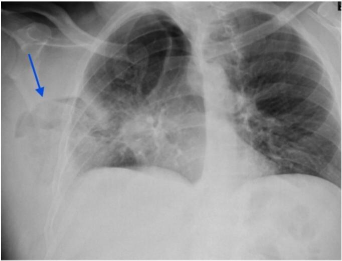
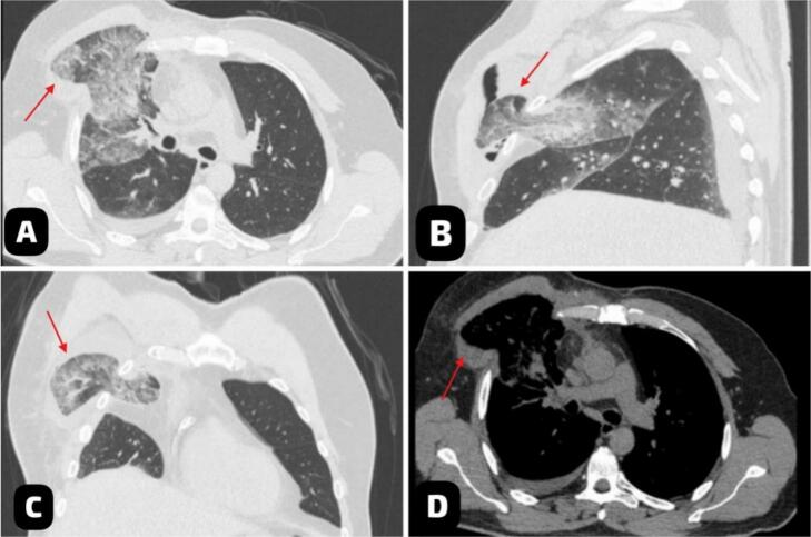
Case presentation: A 57-year-old male with cardiac disease underwent minimally invasive septal myectomy for HOCM. Ten days later, he developed shortness of breath, dry cough, and a right chest mass. Imaging studies, including a chest X-ray revealed a triangular lucency lateral to the right 4th and 5th ribs and non-contrast CT confirmed lung herniation due to an intercostal muscle tear. He underwent surgical repair with rib plating and mesh, leading to significant improvement. He was discharged after seven days and remained asymptomatic at 8-month follow-up.
Discussion: Lung herniation after MICS is rare but serious, caused by intercostal muscle injury. Risk factors include lung conditions, pressure changes, and surgery. Small cases may resolve, but larger ones need surgery. Timely imaging and rib plating with mesh prevented complications and ensured recovery.
Conclusion: Lung herniation should be considered in post-MICS patients with respiratory distress and chest abnormalities. Early diagnosis and surgery are crucial to prevent complications. This case emphasizes the importance of prompt recognition and proper surgical management.

 求助内容:
求助内容: 应助结果提醒方式:


