Jonathan E Henning, Taiga Nishihori, Ciara Freeman, Alexander Lazarides, Jinming Song, Davis Kuruvilla, Sebastian Feuerlein, James R Costello
{"title":"进行性多发性骨髓瘤患者的全身MRI。","authors":"Jonathan E Henning, Taiga Nishihori, Ciara Freeman, Alexander Lazarides, Jinming Song, Davis Kuruvilla, Sebastian Feuerlein, James R Costello","doi":"10.1093/bjrcr/uaaf028","DOIUrl":null,"url":null,"abstract":"<p><p>Multiple Myeloma represents a plasma cell disorder that can result in hallmark bony destructive change in addition to other signs of myelomatous disease. Imaging often helps in establishing the diagnosis and staging the patient. There are several different imaging modalities that can provide different levels of insight into the disease extent. We report a unique case of multiple myeloma where the progressive nature of the patient's disease highlights the strengths and limitations of the different imaging approaches. Whole-body MRI represents a noncontrast imaging technique that directly images the bone marrow space, allowing for disease detection that can precede the onset of cortical and trabecular destructive changes. In so doing, whole-body MRI provides a level of insight that far exceeds traditional plain films and even CT. Using the findings from many different imaging modalities (plain films, CT, PET-CT, and whole-body magnetic resonance imaging), we will discuss how imaging can help clinicians to better assess the patient's disease burden and complement the foundations of traditional disease monitoring (serology, histopathology from biopsy, direct clinical exam, and observation).</p>","PeriodicalId":45216,"journal":{"name":"BJR Case Reports","volume":"11 3","pages":"uaaf028"},"PeriodicalIF":0.5000,"publicationDate":"2025-05-08","publicationTypes":"Journal Article","fieldsOfStudy":null,"isOpenAccess":false,"openAccessPdf":"https://www.ncbi.nlm.nih.gov/pmc/articles/PMC12085220/pdf/","citationCount":"0","resultStr":"{\"title\":\"Whole-body MRI for a patient with progressive multiple myeloma.\",\"authors\":\"Jonathan E Henning, Taiga Nishihori, Ciara Freeman, Alexander Lazarides, Jinming Song, Davis Kuruvilla, Sebastian Feuerlein, James R Costello\",\"doi\":\"10.1093/bjrcr/uaaf028\",\"DOIUrl\":null,\"url\":null,\"abstract\":\"<p><p>Multiple Myeloma represents a plasma cell disorder that can result in hallmark bony destructive change in addition to other signs of myelomatous disease. Imaging often helps in establishing the diagnosis and staging the patient. There are several different imaging modalities that can provide different levels of insight into the disease extent. We report a unique case of multiple myeloma where the progressive nature of the patient's disease highlights the strengths and limitations of the different imaging approaches. Whole-body MRI represents a noncontrast imaging technique that directly images the bone marrow space, allowing for disease detection that can precede the onset of cortical and trabecular destructive changes. In so doing, whole-body MRI provides a level of insight that far exceeds traditional plain films and even CT. Using the findings from many different imaging modalities (plain films, CT, PET-CT, and whole-body magnetic resonance imaging), we will discuss how imaging can help clinicians to better assess the patient's disease burden and complement the foundations of traditional disease monitoring (serology, histopathology from biopsy, direct clinical exam, and observation).</p>\",\"PeriodicalId\":45216,\"journal\":{\"name\":\"BJR Case Reports\",\"volume\":\"11 3\",\"pages\":\"uaaf028\"},\"PeriodicalIF\":0.5000,\"publicationDate\":\"2025-05-08\",\"publicationTypes\":\"Journal Article\",\"fieldsOfStudy\":null,\"isOpenAccess\":false,\"openAccessPdf\":\"https://www.ncbi.nlm.nih.gov/pmc/articles/PMC12085220/pdf/\",\"citationCount\":\"0\",\"resultStr\":null,\"platform\":\"Semanticscholar\",\"paperid\":null,\"PeriodicalName\":\"BJR Case Reports\",\"FirstCategoryId\":\"1085\",\"ListUrlMain\":\"https://doi.org/10.1093/bjrcr/uaaf028\",\"RegionNum\":0,\"RegionCategory\":null,\"ArticlePicture\":[],\"TitleCN\":null,\"AbstractTextCN\":null,\"PMCID\":null,\"EPubDate\":\"2025/5/1 0:00:00\",\"PubModel\":\"eCollection\",\"JCR\":\"Q4\",\"JCRName\":\"RADIOLOGY, NUCLEAR MEDICINE & MEDICAL IMAGING\",\"Score\":null,\"Total\":0}","platform":"Semanticscholar","paperid":null,"PeriodicalName":"BJR Case Reports","FirstCategoryId":"1085","ListUrlMain":"https://doi.org/10.1093/bjrcr/uaaf028","RegionNum":0,"RegionCategory":null,"ArticlePicture":[],"TitleCN":null,"AbstractTextCN":null,"PMCID":null,"EPubDate":"2025/5/1 0:00:00","PubModel":"eCollection","JCR":"Q4","JCRName":"RADIOLOGY, NUCLEAR MEDICINE & MEDICAL IMAGING","Score":null,"Total":0}
Whole-body MRI for a patient with progressive multiple myeloma.
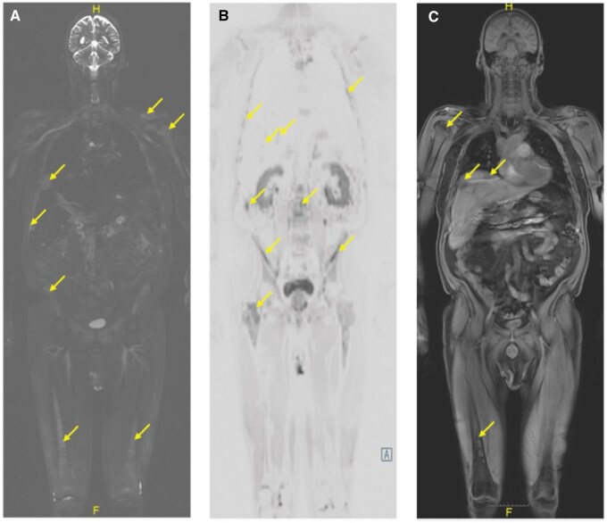
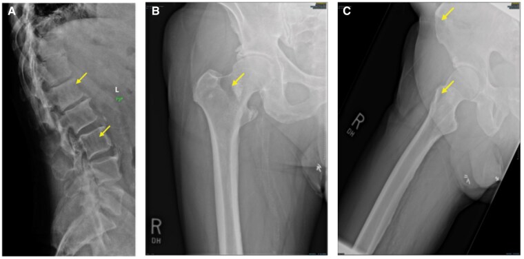
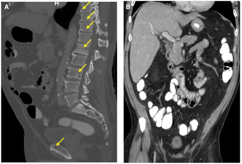
Multiple Myeloma represents a plasma cell disorder that can result in hallmark bony destructive change in addition to other signs of myelomatous disease. Imaging often helps in establishing the diagnosis and staging the patient. There are several different imaging modalities that can provide different levels of insight into the disease extent. We report a unique case of multiple myeloma where the progressive nature of the patient's disease highlights the strengths and limitations of the different imaging approaches. Whole-body MRI represents a noncontrast imaging technique that directly images the bone marrow space, allowing for disease detection that can precede the onset of cortical and trabecular destructive changes. In so doing, whole-body MRI provides a level of insight that far exceeds traditional plain films and even CT. Using the findings from many different imaging modalities (plain films, CT, PET-CT, and whole-body magnetic resonance imaging), we will discuss how imaging can help clinicians to better assess the patient's disease burden and complement the foundations of traditional disease monitoring (serology, histopathology from biopsy, direct clinical exam, and observation).

 求助内容:
求助内容: 应助结果提醒方式:


