利用13c标记葡萄糖代谢的质谱成像鉴定胶质母细胞瘤患者的细胞内在代谢表型
IF 20.8
1区 医学
Q1 ENDOCRINOLOGY & METABOLISM
引用次数: 0
摘要
转录组学研究试图将胶质母细胞瘤(GB)分类为可预测生存率的亚型,并具有不同的治疗脆弱性1,2,3。在这里,我们通过质谱成像鉴定了三种代谢亚型:糖酵解、氧化和糖酵解和氧化的混合,这些质谱成像来自两名输注[U-13C]葡萄糖的GB患者的快速切除肿瘤切片,并通过连续切片的空间转录组学分析。这些表型与微环境特征(包括增殖率、免疫细胞浸润和血管化)无关,当患者来源的细胞在体外生长或作为原位移植的异种移植物生长时,这些表型被保留下来,并且对氧浓度的变化具有很强的适应性,证明了它们的细胞固有性质。显示这些不同代谢表型的细胞所占据的区域的空间范围足够大,可以使用临床适用的代谢成像技术进行检测。这项研究的一个局限性是,它只基于两个病人的肿瘤,尽管是在多个部分,因此代表了一个概念验证研究。本文章由计算机程序翻译,如有差异,请以英文原文为准。
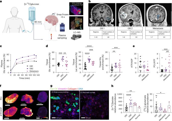

Cell-intrinsic metabolic phenotypes identified in patients with glioblastoma, using mass spectrometry imaging of 13C-labelled glucose metabolism
Transcriptomic studies have attempted to classify glioblastoma (GB) into subtypes that predict survival and have different therapeutic vulnerabilities1–3. Here we identified three metabolic subtypes: glycolytic, oxidative and a mix of glycolytic and oxidative, using mass spectrometry imaging of rapidly excised tumour sections from two patients with GB who were infused with [U-13C]glucose and from spatial transcriptomic analysis of contiguous sections. The phenotypes are not correlated with microenvironmental features, including proliferation rate, immune cell infiltration and vascularization, are retained when patient-derived cells are grown in vitro or as orthotopically implanted xenografts and are robust to changes in oxygen concentration, demonstrating their cell-intrinsic nature. The spatial extent of the regions occupied by cells displaying these distinct metabolic phenotypes is large enough to be detected using clinically applicable metabolic imaging techniques. A limitation of the study is that it is based on only two patient tumours, albeit on multiple sections, and therefore represents a proof-of-concept study. Tsyben et al. study the spatial distribution of labelled metabolites in glioblastoma samples from patients infused with 13C-glucose, using mass spectrometry imaging.
求助全文
通过发布文献求助,成功后即可免费获取论文全文。
去求助
来源期刊
Nature metabolism
ENDOCRINOLOGY & METABOLISM-
CiteScore
27.50
自引率
2.40%
发文量
170
期刊介绍:
Nature Metabolism is a peer-reviewed scientific journal that covers a broad range of topics in metabolism research. It aims to advance the understanding of metabolic and homeostatic processes at a cellular and physiological level. The journal publishes research from various fields, including fundamental cell biology, basic biomedical and translational research, and integrative physiology. It focuses on how cellular metabolism affects cellular function, the physiology and homeostasis of organs and tissues, and the regulation of organismal energy homeostasis. It also investigates the molecular pathophysiology of metabolic diseases such as diabetes and obesity, as well as their treatment. Nature Metabolism follows the standards of other Nature-branded journals, with a dedicated team of professional editors, rigorous peer-review process, high standards of copy-editing and production, swift publication, and editorial independence. The journal has a high impact factor, has a certain influence in the international area, and is deeply concerned and cited by the majority of scholars.
 求助内容:
求助内容: 应助结果提醒方式:


