Wael Hussein, Ahmed Younis, Ahmad Elrawdy, Mohamed ElSholkamy
{"title":"评估引导上颌外侧窦提升术与同时种植体放置立体骨成像手术指南:一项随机对照临床研究。","authors":"Wael Hussein, Ahmed Younis, Ahmad Elrawdy, Mohamed ElSholkamy","doi":"10.1007/s10006-025-01399-3","DOIUrl":null,"url":null,"abstract":"<p><strong>Aim: </strong>The aim of this study is to assess the efficacy of the stereolithographic surgical guide in reducing intraoperative and postoperative complication during lateral sinus lift operation.</p><p><strong>Materials and methods: </strong>A parallel randomized controlled prospective clinical study was conducted on fourteen patients requiring thirty dental implants in the posterior maxilla and diagnosed with reduced vertical bone height. Lateral Maxillary Sinus Lift procedure with simultaneous implant placement was performed to all patients. Stereolithographic surgical guides for lateral window osteotomy and implant drilling and placement were used in the study group, while lateral osteotomy and implant drilling and placement was done freehand in the control group. A cone beam computed tomography was taken immediately and six months post-sinus lifting. Intraoperative and postoperative complications were assessed, pain and edema were assessed using visual analogue scale and vertical bone was assessed using fusion module of cone beam computed tomography.</p><p><strong>Results: </strong>All dental implants demonstrated high survival rates with no statistically significant difference observed in intraoperative or postoperative complications. In terms of new vertical bone gain, both groups exhibited satisfactory and successful outcomes. Concerning pain, there was no statistically significant difference between the two groups except after two days, the study group showed statistically significantly lower pain score than the control group. While regarding the severity of edema, the study group showed statistically significantly higher prevalence of moderate and severe edema than control group which showed higher prevalence of mild edema.</p><p><strong>Conclusion: </strong>According to the current study it has been concluded that there was no remarkable difference between the outcomes of both methods. The study protocol and its consent form were approved by the ethical committee of Suez Canal University (No.432/2021); and registered retrospectively on 23 April 2024 on PACTR (PACTR20240875463218) (pactr.samrc.ac.za/TrialDisplay.aspx?TrialID = 30442).</p>","PeriodicalId":47251,"journal":{"name":"Oral and Maxillofacial Surgery-Heidelberg","volume":"29 1","pages":"102"},"PeriodicalIF":1.8000,"publicationDate":"2025-05-17","publicationTypes":"Journal Article","fieldsOfStudy":null,"isOpenAccess":false,"openAccessPdf":"https://www.ncbi.nlm.nih.gov/pmc/articles/PMC12085366/pdf/","citationCount":"0","resultStr":"{\"title\":\"Assessment of guided lateral maxillary sinus lift procedure with simultaneous implant placement using stereolithographic surgical guide: a randomized controlled clinical study.\",\"authors\":\"Wael Hussein, Ahmed Younis, Ahmad Elrawdy, Mohamed ElSholkamy\",\"doi\":\"10.1007/s10006-025-01399-3\",\"DOIUrl\":null,\"url\":null,\"abstract\":\"<p><strong>Aim: </strong>The aim of this study is to assess the efficacy of the stereolithographic surgical guide in reducing intraoperative and postoperative complication during lateral sinus lift operation.</p><p><strong>Materials and methods: </strong>A parallel randomized controlled prospective clinical study was conducted on fourteen patients requiring thirty dental implants in the posterior maxilla and diagnosed with reduced vertical bone height. Lateral Maxillary Sinus Lift procedure with simultaneous implant placement was performed to all patients. Stereolithographic surgical guides for lateral window osteotomy and implant drilling and placement were used in the study group, while lateral osteotomy and implant drilling and placement was done freehand in the control group. A cone beam computed tomography was taken immediately and six months post-sinus lifting. Intraoperative and postoperative complications were assessed, pain and edema were assessed using visual analogue scale and vertical bone was assessed using fusion module of cone beam computed tomography.</p><p><strong>Results: </strong>All dental implants demonstrated high survival rates with no statistically significant difference observed in intraoperative or postoperative complications. In terms of new vertical bone gain, both groups exhibited satisfactory and successful outcomes. Concerning pain, there was no statistically significant difference between the two groups except after two days, the study group showed statistically significantly lower pain score than the control group. While regarding the severity of edema, the study group showed statistically significantly higher prevalence of moderate and severe edema than control group which showed higher prevalence of mild edema.</p><p><strong>Conclusion: </strong>According to the current study it has been concluded that there was no remarkable difference between the outcomes of both methods. The study protocol and its consent form were approved by the ethical committee of Suez Canal University (No.432/2021); and registered retrospectively on 23 April 2024 on PACTR (PACTR20240875463218) (pactr.samrc.ac.za/TrialDisplay.aspx?TrialID = 30442).</p>\",\"PeriodicalId\":47251,\"journal\":{\"name\":\"Oral and Maxillofacial Surgery-Heidelberg\",\"volume\":\"29 1\",\"pages\":\"102\"},\"PeriodicalIF\":1.8000,\"publicationDate\":\"2025-05-17\",\"publicationTypes\":\"Journal Article\",\"fieldsOfStudy\":null,\"isOpenAccess\":false,\"openAccessPdf\":\"https://www.ncbi.nlm.nih.gov/pmc/articles/PMC12085366/pdf/\",\"citationCount\":\"0\",\"resultStr\":null,\"platform\":\"Semanticscholar\",\"paperid\":null,\"PeriodicalName\":\"Oral and Maxillofacial Surgery-Heidelberg\",\"FirstCategoryId\":\"1085\",\"ListUrlMain\":\"https://doi.org/10.1007/s10006-025-01399-3\",\"RegionNum\":0,\"RegionCategory\":null,\"ArticlePicture\":[],\"TitleCN\":null,\"AbstractTextCN\":null,\"PMCID\":null,\"EPubDate\":\"\",\"PubModel\":\"\",\"JCR\":\"Q3\",\"JCRName\":\"DENTISTRY, ORAL SURGERY & MEDICINE\",\"Score\":null,\"Total\":0}","platform":"Semanticscholar","paperid":null,"PeriodicalName":"Oral and Maxillofacial Surgery-Heidelberg","FirstCategoryId":"1085","ListUrlMain":"https://doi.org/10.1007/s10006-025-01399-3","RegionNum":0,"RegionCategory":null,"ArticlePicture":[],"TitleCN":null,"AbstractTextCN":null,"PMCID":null,"EPubDate":"","PubModel":"","JCR":"Q3","JCRName":"DENTISTRY, ORAL SURGERY & MEDICINE","Score":null,"Total":0}
Assessment of guided lateral maxillary sinus lift procedure with simultaneous implant placement using stereolithographic surgical guide: a randomized controlled clinical study.
Aim: The aim of this study is to assess the efficacy of the stereolithographic surgical guide in reducing intraoperative and postoperative complication during lateral sinus lift operation.
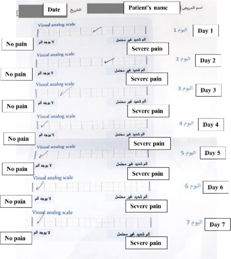
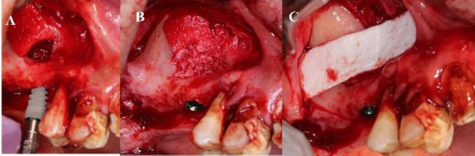
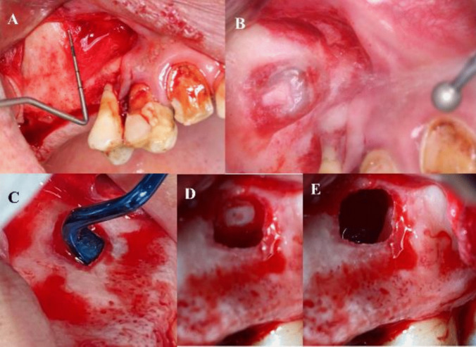
Materials and methods: A parallel randomized controlled prospective clinical study was conducted on fourteen patients requiring thirty dental implants in the posterior maxilla and diagnosed with reduced vertical bone height. Lateral Maxillary Sinus Lift procedure with simultaneous implant placement was performed to all patients. Stereolithographic surgical guides for lateral window osteotomy and implant drilling and placement were used in the study group, while lateral osteotomy and implant drilling and placement was done freehand in the control group. A cone beam computed tomography was taken immediately and six months post-sinus lifting. Intraoperative and postoperative complications were assessed, pain and edema were assessed using visual analogue scale and vertical bone was assessed using fusion module of cone beam computed tomography.
Results: All dental implants demonstrated high survival rates with no statistically significant difference observed in intraoperative or postoperative complications. In terms of new vertical bone gain, both groups exhibited satisfactory and successful outcomes. Concerning pain, there was no statistically significant difference between the two groups except after two days, the study group showed statistically significantly lower pain score than the control group. While regarding the severity of edema, the study group showed statistically significantly higher prevalence of moderate and severe edema than control group which showed higher prevalence of mild edema.
Conclusion: According to the current study it has been concluded that there was no remarkable difference between the outcomes of both methods. The study protocol and its consent form were approved by the ethical committee of Suez Canal University (No.432/2021); and registered retrospectively on 23 April 2024 on PACTR (PACTR20240875463218) (pactr.samrc.ac.za/TrialDisplay.aspx?TrialID = 30442).
期刊介绍:
Oral & Maxillofacial Surgery founded as Mund-, Kiefer- und Gesichtschirurgie is a peer-reviewed online journal. It is designed for clinicians as well as researchers.The quarterly journal offers comprehensive coverage of new techniques, important developments and innovative ideas in oral and maxillofacial surgery and interdisciplinary aspects of cranial, facial and oral diseases and their management. The journal publishes papers of the highest scientific merit and widest possible scope on work in oral and maxillofacial surgery as well as supporting specialties. Practice-oriented articles help improve the methods used in oral and maxillofacial surgery.Every aspect of oral and maxillofacial surgery is fully covered through a range of invited review articles, clinical and research articles, technical notes, abstracts, and case reports. Specific topics are: aesthetic facial surgery, clinical pathology, computer-assisted surgery, congenital and craniofacial deformities, dentoalveolar surgery, head and neck oncology, implant dentistry, oral medicine, orthognathic surgery, reconstructive surgery, skull base surgery, TMJ and trauma.Time-limited reviewing and electronic processing allow to publish articles as fast as possible. Accepted articles are rapidly accessible online.Clinical studies submitted for publication have to include a declaration that they have been approved by an ethical committee according to the World Medical Association Declaration of Helsinki 1964 (last amendment during the 52nd World Medical Association General Assembly, Edinburgh, Scotland, October 2000). Experimental animal studies have to be carried out according to the principles of laboratory animal care (NIH publication No 86-23, revised 1985).
 求助内容:
求助内容: 应助结果提醒方式:


