三种埃及蛇:白天活动的sibilans和schokari以及白天和夜间活动的Spalerosophis diadema的视网膜显微结构-视觉日常活动关系的新见解

IF 1.5 3区 生物学 Q2 ZOOLOGY
Doaa Gewily , Ramadan Kandyel , Fatma Madkour , Mohamed Abumandour , Om Prakash Choudhary , Aya H. Tantawy , Walaa Shalaby
{"title":"三种埃及蛇:白天活动的sibilans和schokari以及白天和夜间活动的Spalerosophis diadema的视网膜显微结构-视觉日常活动关系的新见解","authors":"Doaa Gewily ,&nbsp;Ramadan Kandyel ,&nbsp;Fatma Madkour ,&nbsp;Mohamed Abumandour ,&nbsp;Om Prakash Choudhary ,&nbsp;Aya H. Tantawy ,&nbsp;Walaa Shalaby","doi":"10.1016/j.jcz.2025.05.003","DOIUrl":null,"url":null,"abstract":"<div><h3>Background</h3><div>The retina is adapted to the visual activity of vertebrates, and this study attempts to clarify the retinal morphological features between three snakes with different visual activities.</div></div><div><h3>Methods</h3><div>The retina of three Egyptian snakes of different visual activities (circadian activity)—diurnal <em>Psammophis sibilans</em>, diurnal <em>Psammophis schokari</em>, and frequently diurnal <em>active</em> and <em>little</em> nocturnal active <em>Spalerosophis diadema</em>—have been investigated by light and transmission electron microscopy.</div></div><div><h3>Results</h3><div>The study examined the morphology of photoreceptors in three snakes and their retinal structure to understand their adaptation to different life modes. Our results showed that diurnal snakes have cone-type photoreceptors, while nocturnal snakes have rod-type photoreceptors. The vascular retina consists of two layers: the outer pigmented epithelium and the inner neural layer, which consists of nine layers as described in other snakes. The pigmented epithelium consists of a single cellular layer that extends externally to the photoreceptor layer. The photoreceptor cell layer in <em>P. sibilans</em> comprised single and double cones, while <em>P. schokari</em> and <em>S. diadema</em> had only a single cone, but in <em>P. schokari,</em> this single cone had two types (small and large). The photoreceptor layer in <em>S. diadema</em> is thicker than in <em>P. sibilans</em> and <em>P. schokari</em>. There are a lot of differences between the layer thicknesses of the studied snakes.</div></div><div><h3>Conclusion</h3><div>Our findings revealed that the retinal microstructure in the three snakes was linked to their visual activity.</div></div>","PeriodicalId":49332,"journal":{"name":"Zoologischer Anzeiger","volume":"316 ","pages":"Pages 309-319"},"PeriodicalIF":1.5000,"publicationDate":"2025-05-01","publicationTypes":"Journal Article","fieldsOfStudy":null,"isOpenAccess":false,"openAccessPdf":"","citationCount":"0","resultStr":"{\"title\":\"New insights into the comparative retinal microstructure-visual daily activity relationship among three Egyptian snakes: Diurnal Psammophis sibilans and Psammophis schokari, and both diurnal and nocturnal Spalerosophis diadema\",\"authors\":\"Doaa Gewily ,&nbsp;Ramadan Kandyel ,&nbsp;Fatma Madkour ,&nbsp;Mohamed Abumandour ,&nbsp;Om Prakash Choudhary ,&nbsp;Aya H. Tantawy ,&nbsp;Walaa Shalaby\",\"doi\":\"10.1016/j.jcz.2025.05.003\",\"DOIUrl\":null,\"url\":null,\"abstract\":\"<div><h3>Background</h3><div>The retina is adapted to the visual activity of vertebrates, and this study attempts to clarify the retinal morphological features between three snakes with different visual activities.</div></div><div><h3>Methods</h3><div>The retina of three Egyptian snakes of different visual activities (circadian activity)—diurnal <em>Psammophis sibilans</em>, diurnal <em>Psammophis schokari</em>, and frequently diurnal <em>active</em> and <em>little</em> nocturnal active <em>Spalerosophis diadema</em>—have been investigated by light and transmission electron microscopy.</div></div><div><h3>Results</h3><div>The study examined the morphology of photoreceptors in three snakes and their retinal structure to understand their adaptation to different life modes. Our results showed that diurnal snakes have cone-type photoreceptors, while nocturnal snakes have rod-type photoreceptors. The vascular retina consists of two layers: the outer pigmented epithelium and the inner neural layer, which consists of nine layers as described in other snakes. The pigmented epithelium consists of a single cellular layer that extends externally to the photoreceptor layer. The photoreceptor cell layer in <em>P. sibilans</em> comprised single and double cones, while <em>P. schokari</em> and <em>S. diadema</em> had only a single cone, but in <em>P. schokari,</em> this single cone had two types (small and large). The photoreceptor layer in <em>S. diadema</em> is thicker than in <em>P. sibilans</em> and <em>P. schokari</em>. There are a lot of differences between the layer thicknesses of the studied snakes.</div></div><div><h3>Conclusion</h3><div>Our findings revealed that the retinal microstructure in the three snakes was linked to their visual activity.</div></div>\",\"PeriodicalId\":49332,\"journal\":{\"name\":\"Zoologischer Anzeiger\",\"volume\":\"316 \",\"pages\":\"Pages 309-319\"},\"PeriodicalIF\":1.5000,\"publicationDate\":\"2025-05-01\",\"publicationTypes\":\"Journal Article\",\"fieldsOfStudy\":null,\"isOpenAccess\":false,\"openAccessPdf\":\"\",\"citationCount\":\"0\",\"resultStr\":null,\"platform\":\"Semanticscholar\",\"paperid\":null,\"PeriodicalName\":\"Zoologischer Anzeiger\",\"FirstCategoryId\":\"99\",\"ListUrlMain\":\"https://www.sciencedirect.com/science/article/pii/S0044523125000476\",\"RegionNum\":3,\"RegionCategory\":\"生物学\",\"ArticlePicture\":[],\"TitleCN\":null,\"AbstractTextCN\":null,\"PMCID\":null,\"EPubDate\":\"\",\"PubModel\":\"\",\"JCR\":\"Q2\",\"JCRName\":\"ZOOLOGY\",\"Score\":null,\"Total\":0}","platform":"Semanticscholar","paperid":null,"PeriodicalName":"Zoologischer Anzeiger","FirstCategoryId":"99","ListUrlMain":"https://www.sciencedirect.com/science/article/pii/S0044523125000476","RegionNum":3,"RegionCategory":"生物学","ArticlePicture":[],"TitleCN":null,"AbstractTextCN":null,"PMCID":null,"EPubDate":"","PubModel":"","JCR":"Q2","JCRName":"ZOOLOGY","Score":null,"Total":0}
引用次数: 0

摘要

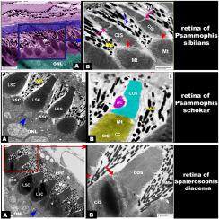
视网膜适应脊椎动物的视觉活动,本研究试图阐明三种不同视觉活动蛇的视网膜形态特征。方法采用光镜和透射电子显微镜观察了三种不同视觉活动(昼夜活动)的埃及蛇——白天活动的sibilans、白天活动的schokari和夜间活动频繁的diadeaspalerosophis的视网膜。结果研究了三种蛇的光感受器形态和视网膜结构,以了解它们对不同生活方式的适应。结果表明,白天活动的蛇具有锥型光感受器,而夜间活动的蛇具有杆状光感受器。血管视网膜由两层组成:外层色素上皮和内层神经层,内层神经层由其他蛇的九层组成。色素上皮由单个细胞层组成,向外延伸至感光层。sibilans的感光细胞层由单和双锥体组成,而p.s okari和s.s diadema只有一个锥体,但在p.s okari中,这个单锥体有小和大两种类型。棘球绦虫的感光层比棘球绦虫和schokari棘球绦虫更厚。被研究的蛇的层厚度有很多不同。结论三种蛇的视网膜结构与它们的视觉活动有关。
本文章由计算机程序翻译,如有差异,请以英文原文为准。

New insights into the comparative retinal microstructure-visual daily activity relationship among three Egyptian snakes: Diurnal Psammophis sibilans and Psammophis schokari, and both diurnal and nocturnal Spalerosophis diadema

New insights into the comparative retinal microstructure-visual daily activity relationship among three Egyptian snakes: Diurnal Psammophis sibilans and Psammophis schokari, and both diurnal and nocturnal Spalerosophis diadema

Background

The retina is adapted to the visual activity of vertebrates, and this study attempts to clarify the retinal morphological features between three snakes with different visual activities.

Methods

The retina of three Egyptian snakes of different visual activities (circadian activity)—diurnal Psammophis sibilans, diurnal Psammophis schokari, and frequently diurnal active and little nocturnal active Spalerosophis diadema—have been investigated by light and transmission electron microscopy.

Results

The study examined the morphology of photoreceptors in three snakes and their retinal structure to understand their adaptation to different life modes. Our results showed that diurnal snakes have cone-type photoreceptors, while nocturnal snakes have rod-type photoreceptors. The vascular retina consists of two layers: the outer pigmented epithelium and the inner neural layer, which consists of nine layers as described in other snakes. The pigmented epithelium consists of a single cellular layer that extends externally to the photoreceptor layer. The photoreceptor cell layer in P. sibilans comprised single and double cones, while P. schokari and S. diadema had only a single cone, but in P. schokari, this single cone had two types (small and large). The photoreceptor layer in S. diadema is thicker than in P. sibilans and P. schokari. There are a lot of differences between the layer thicknesses of the studied snakes.

Conclusion

Our findings revealed that the retinal microstructure in the three snakes was linked to their visual activity.
求助全文
通过发布文献求助,成功后即可免费获取论文全文。 去求助
来源期刊
Zoologischer Anzeiger
Zoologischer Anzeiger 生物-动物学
CiteScore
2.80
自引率
7.10%
发文量
75
审稿时长
>12 weeks
期刊介绍: Zoologischer Anzeiger - A Journal of Comparative Zoology is devoted to comparative zoology with a special emphasis on morphology, systematics, biogeography, and evolutionary biology targeting all metazoans, both modern and extinct. We also consider taxonomic submissions addressing a broader systematic and/or evolutionary context. The overall aim of the journal is to contribute to our understanding of the organismic world from an evolutionary perspective. The journal Zoologischer Anzeiger invites suggestions for special issues. Interested parties may contact one of the editors.
×
引用
GB/T 7714-2015
复制
MLA
复制
APA
复制
导出至
BibTeX EndNote RefMan NoteFirst NoteExpress
×
提示
您的信息不完整,为了账户安全,请先补充。
现在去补充
×
提示
您因"违规操作"
具体请查看互助需知
我知道了
×
提示
确定
请完成安全验证×
copy
已复制链接
快去分享给好友吧!
我知道了
右上角分享
点击右上角分享
0
联系我们:info@booksci.cn Book学术提供免费学术资源搜索服务,方便国内外学者检索中英文文献。致力于提供最便捷和优质的服务体验。 Copyright © 2023 布克学术 All rights reserved.
京ICP备2023020795号-1
ghs 京公网安备 11010802042870号
Book学术文献互助
Book学术文献互助群
群 号:604180095
Book学术官方微信