{"title":"镰状细胞性贫血患者视网膜和脉络膜微血管和超微结构的改变。","authors":"Oğuzhan Oruz, Süheyl Asma, Aysel Pelit, Çiğdem Gereklioğlu, Selçuk Sızmaz, Astan İbayev, Handan Canan, Osman Şahin, Mutlu Kasar, Can Boğa, Caner İncekaş","doi":"10.4274/tjo.galenos.2025.97792","DOIUrl":null,"url":null,"abstract":"<p><strong>Objectives: </strong>To determine the microvascular changes of the retina and choroid in sickle cell anemia (SCA) patients and to investigate the relationship between the severity of sickle cell retinopathy and sickle cell maculopathy (SCM).</p><p><strong>Materials and methods: </strong>In this cross-sectional study, 78 eyes of 39 patients with SCA were included in the patient group and 68 eyes of 34 healthy participants were included in the control group. Differences in foveal avascular zone (FAZ), retinal and subfoveal choroidal thickness (SFCT), and choroidal vascularity index (CVI) between the patient group and the control group were evaluated by swept source optical coherence tomography (OCT) and OCT angiography (OCTA) imaging. In addition, systemic and biological parameters were compared in patients with and without SCM.</p><p><strong>Results: </strong>SCM was detected in 16 eyes of 8 patients. Proliferative sickle cell retinopathy (PSCR) was present in 10 patients. In logistic regression analysis, PSCR was found to be a risk factor for the development of SCM (p=0.015, odds ratio: 17.25, 95% confidence interval: 1.73-172.02). The temporal inner retinal layers were significantly thinner in the patient group compared to the control group. The patient group also exhibited significantly greater FAZ enlargement in both the superficial and deep capillary plexus when compared with the control group (p<0.001 for both). CVI was higher in the control group than in the patient group (p<0.001). SFCT was significantly thinner in the patient group (p=0.013). There was no significant difference between patients with and without SCM in terms of FAZ enlargement, CVI values, or systemic and biological factors.</p><p><strong>Conclusion: </strong>In our study, PSCR was found to be a risk factor for the development of SCM. OCT and OCTA provide valuable information about microvascular changes in the retina and choroid in patients with SCM. Structural changes demonstrated by OCTA before the development of SCM are very important for follow-up and treatment in terms of visual prognosis of patients.</p>","PeriodicalId":23373,"journal":{"name":"Turkish Journal of Ophthalmology","volume":"55 2","pages":"74-81"},"PeriodicalIF":0.0000,"publicationDate":"2025-04-24","publicationTypes":"Journal Article","fieldsOfStudy":null,"isOpenAccess":false,"openAccessPdf":"https://www.ncbi.nlm.nih.gov/pmc/articles/PMC12035012/pdf/","citationCount":"0","resultStr":"{\"title\":\"Microvascular and Ultrastructural Changes of the Retina and Choroid in Patients with Sickle Cell Anemia.\",\"authors\":\"Oğuzhan Oruz, Süheyl Asma, Aysel Pelit, Çiğdem Gereklioğlu, Selçuk Sızmaz, Astan İbayev, Handan Canan, Osman Şahin, Mutlu Kasar, Can Boğa, Caner İncekaş\",\"doi\":\"10.4274/tjo.galenos.2025.97792\",\"DOIUrl\":null,\"url\":null,\"abstract\":\"<p><strong>Objectives: </strong>To determine the microvascular changes of the retina and choroid in sickle cell anemia (SCA) patients and to investigate the relationship between the severity of sickle cell retinopathy and sickle cell maculopathy (SCM).</p><p><strong>Materials and methods: </strong>In this cross-sectional study, 78 eyes of 39 patients with SCA were included in the patient group and 68 eyes of 34 healthy participants were included in the control group. Differences in foveal avascular zone (FAZ), retinal and subfoveal choroidal thickness (SFCT), and choroidal vascularity index (CVI) between the patient group and the control group were evaluated by swept source optical coherence tomography (OCT) and OCT angiography (OCTA) imaging. In addition, systemic and biological parameters were compared in patients with and without SCM.</p><p><strong>Results: </strong>SCM was detected in 16 eyes of 8 patients. Proliferative sickle cell retinopathy (PSCR) was present in 10 patients. In logistic regression analysis, PSCR was found to be a risk factor for the development of SCM (p=0.015, odds ratio: 17.25, 95% confidence interval: 1.73-172.02). The temporal inner retinal layers were significantly thinner in the patient group compared to the control group. The patient group also exhibited significantly greater FAZ enlargement in both the superficial and deep capillary plexus when compared with the control group (p<0.001 for both). CVI was higher in the control group than in the patient group (p<0.001). SFCT was significantly thinner in the patient group (p=0.013). There was no significant difference between patients with and without SCM in terms of FAZ enlargement, CVI values, or systemic and biological factors.</p><p><strong>Conclusion: </strong>In our study, PSCR was found to be a risk factor for the development of SCM. OCT and OCTA provide valuable information about microvascular changes in the retina and choroid in patients with SCM. Structural changes demonstrated by OCTA before the development of SCM are very important for follow-up and treatment in terms of visual prognosis of patients.</p>\",\"PeriodicalId\":23373,\"journal\":{\"name\":\"Turkish Journal of Ophthalmology\",\"volume\":\"55 2\",\"pages\":\"74-81\"},\"PeriodicalIF\":0.0000,\"publicationDate\":\"2025-04-24\",\"publicationTypes\":\"Journal Article\",\"fieldsOfStudy\":null,\"isOpenAccess\":false,\"openAccessPdf\":\"https://www.ncbi.nlm.nih.gov/pmc/articles/PMC12035012/pdf/\",\"citationCount\":\"0\",\"resultStr\":null,\"platform\":\"Semanticscholar\",\"paperid\":null,\"PeriodicalName\":\"Turkish Journal of Ophthalmology\",\"FirstCategoryId\":\"1085\",\"ListUrlMain\":\"https://doi.org/10.4274/tjo.galenos.2025.97792\",\"RegionNum\":0,\"RegionCategory\":null,\"ArticlePicture\":[],\"TitleCN\":null,\"AbstractTextCN\":null,\"PMCID\":null,\"EPubDate\":\"\",\"PubModel\":\"\",\"JCR\":\"Q3\",\"JCRName\":\"Medicine\",\"Score\":null,\"Total\":0}","platform":"Semanticscholar","paperid":null,"PeriodicalName":"Turkish Journal of Ophthalmology","FirstCategoryId":"1085","ListUrlMain":"https://doi.org/10.4274/tjo.galenos.2025.97792","RegionNum":0,"RegionCategory":null,"ArticlePicture":[],"TitleCN":null,"AbstractTextCN":null,"PMCID":null,"EPubDate":"","PubModel":"","JCR":"Q3","JCRName":"Medicine","Score":null,"Total":0}
Microvascular and Ultrastructural Changes of the Retina and Choroid in Patients with Sickle Cell Anemia.
Objectives: To determine the microvascular changes of the retina and choroid in sickle cell anemia (SCA) patients and to investigate the relationship between the severity of sickle cell retinopathy and sickle cell maculopathy (SCM).
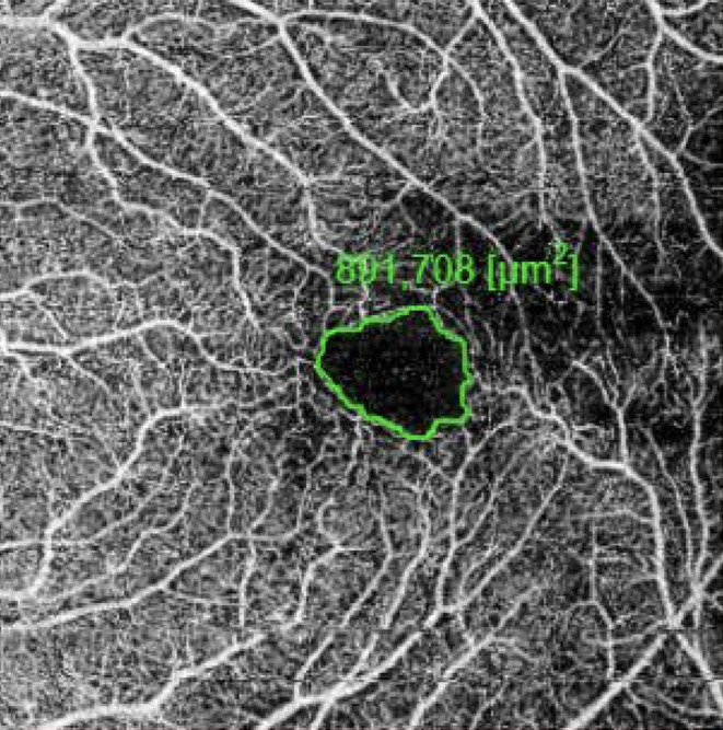
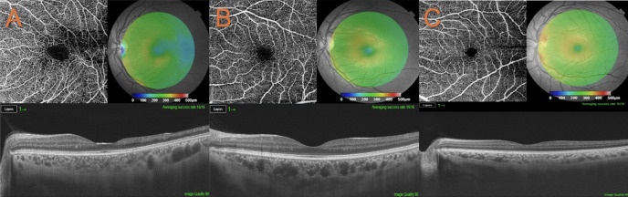
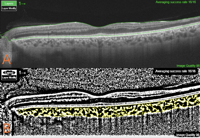
Materials and methods: In this cross-sectional study, 78 eyes of 39 patients with SCA were included in the patient group and 68 eyes of 34 healthy participants were included in the control group. Differences in foveal avascular zone (FAZ), retinal and subfoveal choroidal thickness (SFCT), and choroidal vascularity index (CVI) between the patient group and the control group were evaluated by swept source optical coherence tomography (OCT) and OCT angiography (OCTA) imaging. In addition, systemic and biological parameters were compared in patients with and without SCM.
Results: SCM was detected in 16 eyes of 8 patients. Proliferative sickle cell retinopathy (PSCR) was present in 10 patients. In logistic regression analysis, PSCR was found to be a risk factor for the development of SCM (p=0.015, odds ratio: 17.25, 95% confidence interval: 1.73-172.02). The temporal inner retinal layers were significantly thinner in the patient group compared to the control group. The patient group also exhibited significantly greater FAZ enlargement in both the superficial and deep capillary plexus when compared with the control group (p<0.001 for both). CVI was higher in the control group than in the patient group (p<0.001). SFCT was significantly thinner in the patient group (p=0.013). There was no significant difference between patients with and without SCM in terms of FAZ enlargement, CVI values, or systemic and biological factors.
Conclusion: In our study, PSCR was found to be a risk factor for the development of SCM. OCT and OCTA provide valuable information about microvascular changes in the retina and choroid in patients with SCM. Structural changes demonstrated by OCTA before the development of SCM are very important for follow-up and treatment in terms of visual prognosis of patients.
期刊介绍:
The Turkish Journal of Ophthalmology (TJO) is the only scientific periodical publication of the Turkish Ophthalmological Association and has been published since January 1929. In its early years, the journal was published in Turkish and French. Although there were temporary interruptions in the publication of the journal due to various challenges, the Turkish Journal of Ophthalmology has been published continually from 1971 to the present. The target audience includes specialists and physicians in training in ophthalmology in all relevant disciplines.

 求助内容:
求助内容: 应助结果提醒方式:


