{"title":"一例罕见的成年女性成熟髓内囊性畸胎瘤:1例报告并文献复习。","authors":"Qinyang Zhang, Xuepeng Liu, Haijun Li","doi":"10.5152/j.aott.2025.24039","DOIUrl":null,"url":null,"abstract":"<p><p>A 51-year-old female with a 10-year history of progressive low back pain presented with a 9 × 15 mm circular tumor adjacent to L1 on preoperative magnetic resonance imaging. The lesion was successfully removed by T12-L2 laminectomy and nail-rod fixation. Histopathological examination diagnosedmature intramedullary cystic teratoma. At 6-month follow-up, right lower limb numbness and pain were reduced.</p>","PeriodicalId":93854,"journal":{"name":"Acta orthopaedica et traumatologica turcica","volume":"59 2","pages":"129-132"},"PeriodicalIF":1.0000,"publicationDate":"2025-04-29","publicationTypes":"Journal Article","fieldsOfStudy":null,"isOpenAccess":false,"openAccessPdf":"https://www.ncbi.nlm.nih.gov/pmc/articles/PMC12070423/pdf/","citationCount":"0","resultStr":"{\"title\":\"A rare case of a mature intramedullary cystic teratoma in an adult female: A case report and literature review.\",\"authors\":\"Qinyang Zhang, Xuepeng Liu, Haijun Li\",\"doi\":\"10.5152/j.aott.2025.24039\",\"DOIUrl\":null,\"url\":null,\"abstract\":\"<p><p>A 51-year-old female with a 10-year history of progressive low back pain presented with a 9 × 15 mm circular tumor adjacent to L1 on preoperative magnetic resonance imaging. The lesion was successfully removed by T12-L2 laminectomy and nail-rod fixation. Histopathological examination diagnosedmature intramedullary cystic teratoma. At 6-month follow-up, right lower limb numbness and pain were reduced.</p>\",\"PeriodicalId\":93854,\"journal\":{\"name\":\"Acta orthopaedica et traumatologica turcica\",\"volume\":\"59 2\",\"pages\":\"129-132\"},\"PeriodicalIF\":1.0000,\"publicationDate\":\"2025-04-29\",\"publicationTypes\":\"Journal Article\",\"fieldsOfStudy\":null,\"isOpenAccess\":false,\"openAccessPdf\":\"https://www.ncbi.nlm.nih.gov/pmc/articles/PMC12070423/pdf/\",\"citationCount\":\"0\",\"resultStr\":null,\"platform\":\"Semanticscholar\",\"paperid\":null,\"PeriodicalName\":\"Acta orthopaedica et traumatologica turcica\",\"FirstCategoryId\":\"1085\",\"ListUrlMain\":\"https://doi.org/10.5152/j.aott.2025.24039\",\"RegionNum\":0,\"RegionCategory\":null,\"ArticlePicture\":[],\"TitleCN\":null,\"AbstractTextCN\":null,\"PMCID\":null,\"EPubDate\":\"\",\"PubModel\":\"\",\"JCR\":\"\",\"JCRName\":\"\",\"Score\":null,\"Total\":0}","platform":"Semanticscholar","paperid":null,"PeriodicalName":"Acta orthopaedica et traumatologica turcica","FirstCategoryId":"1085","ListUrlMain":"https://doi.org/10.5152/j.aott.2025.24039","RegionNum":0,"RegionCategory":null,"ArticlePicture":[],"TitleCN":null,"AbstractTextCN":null,"PMCID":null,"EPubDate":"","PubModel":"","JCR":"","JCRName":"","Score":null,"Total":0}
A rare case of a mature intramedullary cystic teratoma in an adult female: A case report and literature review.
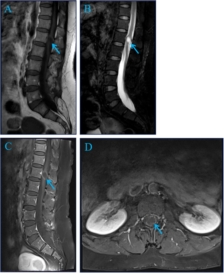
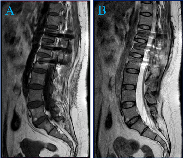
A 51-year-old female with a 10-year history of progressive low back pain presented with a 9 × 15 mm circular tumor adjacent to L1 on preoperative magnetic resonance imaging. The lesion was successfully removed by T12-L2 laminectomy and nail-rod fixation. Histopathological examination diagnosedmature intramedullary cystic teratoma. At 6-month follow-up, right lower limb numbness and pain were reduced.


 求助内容:
求助内容: 应助结果提醒方式:


