{"title":"用机器学习方法和神经影像学改进阿尔茨海默病诊断:案例研究发展。","authors":"Lilia Lazli","doi":"10.2196/60866","DOIUrl":null,"url":null,"abstract":"<p><strong>Background: </strong>Alzheimer disease (AD) is a severe neurological brain disorder. While not curable, earlier detection can help improve symptoms substantially. Machine learning (ML) models are popular and well suited for medical image processing tasks such as computer-aided diagnosis. These techniques can improve the process for an accurate diagnosis of AD.</p><p><strong>Objective: </strong>In this paper, a complete computer-aided diagnosis system for the diagnosis of AD has been presented. We investigate the performance of some of the most used ML techniques for AD detection and classification using neuroimages from the Open Access Series of Imaging Studies (OASIS) and Alzheimer's Disease Neuroimaging Initiative (ADNI) datasets.</p><p><strong>Methods: </strong>The system uses artificial neural networks (ANNs) and support vector machines (SVMs) as classifiers, and dimensionality reduction techniques as feature extractors. To retrieve features from the neuroimages, we used principal component analysis (PCA), linear discriminant analysis, and t-distributed stochastic neighbor embedding. These features are fed into feedforward neural networks (FFNNs) and SVM-based ML classifiers. Furthermore, we applied the vision transformer (ViT)-based ANNs in conjunction with data augmentation to distinguish patients with AD from healthy controls.</p><p><strong>Results: </strong>Experiments were performed on magnetic resonance imaging and positron emission tomography scans. The OASIS dataset included a total of 300 patients, while the ADNI dataset included 231 patients. For OASIS, 90 (30%) patients were healthy and 210 (70%) were severely impaired by AD. Likewise for the ADNI database, a total of 149 (64.5%) patients with AD were detected and 82 (35.5%) patients were used as healthy controls. An important difference was established between healthy patients and patients with AD (P=.02). We examined the effectiveness of the three feature extractors and classifiers using 5-fold cross-validation and confusion matrix-based standard classification metrics, namely, accuracy, sensitivity, specificity, precision, F1-score, and area under the receiver operating characteristic curve (AUROC). Compared with the state-of-the-art performing methods, the success rate was satisfactory for all the created ML models, but SVM and FFNN performed best with the PCA extractor, while the ViT classifier performed best with more data. The data augmentation/ViT approach worked better overall, achieving accuracies of 93.2% (sensitivity=87.2, specificity=90.5, precision=87.6, F1-score=88.7, and AUROC=92) for OASIS and 90.4% (sensitivity=85.4, specificity=88.6, precision=86.9, F1-score=88, and AUROC=90) for ADNI.</p><p><strong>Conclusions: </strong>Effective ML models using neuroimaging data could help physicians working on AD diagnosis and will assist them in prescribing timely treatment to patients with AD. Good results were obtained on the OASIS and ADNI datasets with all the proposed classifiers, namely, SVM, FFNN, and ViTs. However, the results show that the ViT model is much better at predicting AD than the other models when a sufficient amount of data are available to perform the training. This highlights that the data augmentation process could impact the overall performance of the ViT model.</p>","PeriodicalId":73558,"journal":{"name":"JMIRx med","volume":"6 ","pages":"e60866"},"PeriodicalIF":0.0000,"publicationDate":"2025-04-21","publicationTypes":"Journal Article","fieldsOfStudy":null,"isOpenAccess":false,"openAccessPdf":"https://www.ncbi.nlm.nih.gov/pmc/articles/PMC12036548/pdf/","citationCount":"0","resultStr":"{\"title\":\"Improved Alzheimer Disease Diagnosis With a Machine Learning Approach and Neuroimaging: Case Study Development.\",\"authors\":\"Lilia Lazli\",\"doi\":\"10.2196/60866\",\"DOIUrl\":null,\"url\":null,\"abstract\":\"<p><strong>Background: </strong>Alzheimer disease (AD) is a severe neurological brain disorder. While not curable, earlier detection can help improve symptoms substantially. Machine learning (ML) models are popular and well suited for medical image processing tasks such as computer-aided diagnosis. These techniques can improve the process for an accurate diagnosis of AD.</p><p><strong>Objective: </strong>In this paper, a complete computer-aided diagnosis system for the diagnosis of AD has been presented. We investigate the performance of some of the most used ML techniques for AD detection and classification using neuroimages from the Open Access Series of Imaging Studies (OASIS) and Alzheimer's Disease Neuroimaging Initiative (ADNI) datasets.</p><p><strong>Methods: </strong>The system uses artificial neural networks (ANNs) and support vector machines (SVMs) as classifiers, and dimensionality reduction techniques as feature extractors. To retrieve features from the neuroimages, we used principal component analysis (PCA), linear discriminant analysis, and t-distributed stochastic neighbor embedding. These features are fed into feedforward neural networks (FFNNs) and SVM-based ML classifiers. Furthermore, we applied the vision transformer (ViT)-based ANNs in conjunction with data augmentation to distinguish patients with AD from healthy controls.</p><p><strong>Results: </strong>Experiments were performed on magnetic resonance imaging and positron emission tomography scans. The OASIS dataset included a total of 300 patients, while the ADNI dataset included 231 patients. For OASIS, 90 (30%) patients were healthy and 210 (70%) were severely impaired by AD. Likewise for the ADNI database, a total of 149 (64.5%) patients with AD were detected and 82 (35.5%) patients were used as healthy controls. An important difference was established between healthy patients and patients with AD (P=.02). We examined the effectiveness of the three feature extractors and classifiers using 5-fold cross-validation and confusion matrix-based standard classification metrics, namely, accuracy, sensitivity, specificity, precision, F1-score, and area under the receiver operating characteristic curve (AUROC). Compared with the state-of-the-art performing methods, the success rate was satisfactory for all the created ML models, but SVM and FFNN performed best with the PCA extractor, while the ViT classifier performed best with more data. The data augmentation/ViT approach worked better overall, achieving accuracies of 93.2% (sensitivity=87.2, specificity=90.5, precision=87.6, F1-score=88.7, and AUROC=92) for OASIS and 90.4% (sensitivity=85.4, specificity=88.6, precision=86.9, F1-score=88, and AUROC=90) for ADNI.</p><p><strong>Conclusions: </strong>Effective ML models using neuroimaging data could help physicians working on AD diagnosis and will assist them in prescribing timely treatment to patients with AD. Good results were obtained on the OASIS and ADNI datasets with all the proposed classifiers, namely, SVM, FFNN, and ViTs. However, the results show that the ViT model is much better at predicting AD than the other models when a sufficient amount of data are available to perform the training. This highlights that the data augmentation process could impact the overall performance of the ViT model.</p>\",\"PeriodicalId\":73558,\"journal\":{\"name\":\"JMIRx med\",\"volume\":\"6 \",\"pages\":\"e60866\"},\"PeriodicalIF\":0.0000,\"publicationDate\":\"2025-04-21\",\"publicationTypes\":\"Journal Article\",\"fieldsOfStudy\":null,\"isOpenAccess\":false,\"openAccessPdf\":\"https://www.ncbi.nlm.nih.gov/pmc/articles/PMC12036548/pdf/\",\"citationCount\":\"0\",\"resultStr\":null,\"platform\":\"Semanticscholar\",\"paperid\":null,\"PeriodicalName\":\"JMIRx med\",\"FirstCategoryId\":\"1085\",\"ListUrlMain\":\"https://doi.org/10.2196/60866\",\"RegionNum\":0,\"RegionCategory\":null,\"ArticlePicture\":[],\"TitleCN\":null,\"AbstractTextCN\":null,\"PMCID\":null,\"EPubDate\":\"\",\"PubModel\":\"\",\"JCR\":\"\",\"JCRName\":\"\",\"Score\":null,\"Total\":0}","platform":"Semanticscholar","paperid":null,"PeriodicalName":"JMIRx med","FirstCategoryId":"1085","ListUrlMain":"https://doi.org/10.2196/60866","RegionNum":0,"RegionCategory":null,"ArticlePicture":[],"TitleCN":null,"AbstractTextCN":null,"PMCID":null,"EPubDate":"","PubModel":"","JCR":"","JCRName":"","Score":null,"Total":0}
Improved Alzheimer Disease Diagnosis With a Machine Learning Approach and Neuroimaging: Case Study Development.
Background: Alzheimer disease (AD) is a severe neurological brain disorder. While not curable, earlier detection can help improve symptoms substantially. Machine learning (ML) models are popular and well suited for medical image processing tasks such as computer-aided diagnosis. These techniques can improve the process for an accurate diagnosis of AD.
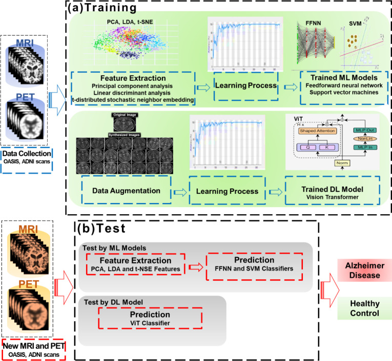
Objective: In this paper, a complete computer-aided diagnosis system for the diagnosis of AD has been presented. We investigate the performance of some of the most used ML techniques for AD detection and classification using neuroimages from the Open Access Series of Imaging Studies (OASIS) and Alzheimer's Disease Neuroimaging Initiative (ADNI) datasets.
Methods: The system uses artificial neural networks (ANNs) and support vector machines (SVMs) as classifiers, and dimensionality reduction techniques as feature extractors. To retrieve features from the neuroimages, we used principal component analysis (PCA), linear discriminant analysis, and t-distributed stochastic neighbor embedding. These features are fed into feedforward neural networks (FFNNs) and SVM-based ML classifiers. Furthermore, we applied the vision transformer (ViT)-based ANNs in conjunction with data augmentation to distinguish patients with AD from healthy controls.
Results: Experiments were performed on magnetic resonance imaging and positron emission tomography scans. The OASIS dataset included a total of 300 patients, while the ADNI dataset included 231 patients. For OASIS, 90 (30%) patients were healthy and 210 (70%) were severely impaired by AD. Likewise for the ADNI database, a total of 149 (64.5%) patients with AD were detected and 82 (35.5%) patients were used as healthy controls. An important difference was established between healthy patients and patients with AD (P=.02). We examined the effectiveness of the three feature extractors and classifiers using 5-fold cross-validation and confusion matrix-based standard classification metrics, namely, accuracy, sensitivity, specificity, precision, F1-score, and area under the receiver operating characteristic curve (AUROC). Compared with the state-of-the-art performing methods, the success rate was satisfactory for all the created ML models, but SVM and FFNN performed best with the PCA extractor, while the ViT classifier performed best with more data. The data augmentation/ViT approach worked better overall, achieving accuracies of 93.2% (sensitivity=87.2, specificity=90.5, precision=87.6, F1-score=88.7, and AUROC=92) for OASIS and 90.4% (sensitivity=85.4, specificity=88.6, precision=86.9, F1-score=88, and AUROC=90) for ADNI.
Conclusions: Effective ML models using neuroimaging data could help physicians working on AD diagnosis and will assist them in prescribing timely treatment to patients with AD. Good results were obtained on the OASIS and ADNI datasets with all the proposed classifiers, namely, SVM, FFNN, and ViTs. However, the results show that the ViT model is much better at predicting AD than the other models when a sufficient amount of data are available to perform the training. This highlights that the data augmentation process could impact the overall performance of the ViT model.

 求助内容:
求助内容: 应助结果提醒方式:


