{"title":"多参数4D-MRI在肝癌放疗中的剂量学研究。","authors":"Sha Li, Xianggao Zhu, Haonan Xiao, Weiwei Liu, Yibao Zhang, Jing Cai, Tian Li, Yanye Lu","doi":"10.1186/s13014-025-02600-3","DOIUrl":null,"url":null,"abstract":"<p><strong>Background: </strong>In radiotherapy, inadequate management of organ motion in liver cancer may lead to inadequate delineation accuracy, resulting in the underdosage of target tissues and overdosage of surrounding normal tissues. To investigate the clinical potential of multi-parametric 4D-MRI in the target delineation and dose accuracy for liver cancer radiotherapy.</p><p><strong>Methods: </strong>Twenty patients receiving radiotherapy for liver cancer were enrolled. Each patient underwent contrast-enhanced planning CT (free-breathing), contrast-enhanced T1-weighted (free-breathing), T2-weighted (gated) 3D-MRI, and low-quality 4D-MRI using the time resolved imaging with interleaved stochastic trajectories volumetric interpolated breath-hold examination (TWIST-VIBE) sequence. A dual-supervised deformation estimation model was used to generate a 4D deformable vector field (4D-DVF) from 4D-MRI data, and the prior images were deformed using this 4D-DVF to generate multi-parametric 4D-MRI. Assisted by 3D-MRI and multi-parametric 4D-MRI, target contours were performed on the planning CT, resulting in the generation of Target_3D and Target_4D. Clinical plans, Plan_3D and Plan_4D, were designed based on these contours respectively. To explore the dosimetric variations resulting from different contours without re-optimization, Plan_3D was directly applied to Target_4D, and Plan_4D was applied to Target_3D to generate Plan_3D' and Plan_4D' respectively. Target volume, contours, dose-volume histograms (DVHs), conformity index (CI), homogeneity index (HI), maximum and mean dose to organ as risks (OARs) were compared and evaluated.</p><p><strong>Results: </strong>Mean volume differences between Target_3D and Target_4D were 2.76 cm<sup>3</sup> (standard deviation [SD] 3.42 cm<sup>3</sup>) in the caudate lobe, 181.54 cm<sup>3</sup> (SD 68.50 cm<sup>3</sup>) in the left hepatic lobe, and 26.08 cm<sup>3</sup> (SD 20.52 cm<sup>3</sup>) in the right hepatic lobe. Mean and SD of CI and HI is 1.02 ± 0.04 and 0.108 ± 0.02 in Plan_3D, 1.02 ± 0.01 and 0.107 ± 0.01 in Plan_4D. There were no statistically significant differences in OAR doses between Plan_3D and Plan_3D', between Plan_4D and Plan_4D'. However, a statistically significant difference in target dose was observed between Plan_3D and Plan_3D' (P = 1.47 × 10⁻⁷) and between Plan_4D and Plan_4D' (P = 0.013). Plan_3D' meets 100% of the prescription dose covering mean 77.89% (SD 10.13%) of the Targeted_4D volume, while Plan_4D' covered mean 94.17% (SD 3.12%) of the Targeted_3D volume.</p><p><strong>Conclusions: </strong>3D image-guided target delineation may be more likely to underestimate target volume and compromise dose coverage, suggesting that using multi-parametric 4D-MRI can provide more precise target contours and enhance target dose coverage.</p>","PeriodicalId":49639,"journal":{"name":"Radiation Oncology","volume":"20 1","pages":"51"},"PeriodicalIF":3.3000,"publicationDate":"2025-04-11","publicationTypes":"Journal Article","fieldsOfStudy":null,"isOpenAccess":false,"openAccessPdf":"https://www.ncbi.nlm.nih.gov/pmc/articles/PMC11987451/pdf/","citationCount":"0","resultStr":"{\"title\":\"Dosimetric investigation of multi-parametric 4D-MRI for radiotherapy in liver cancer.\",\"authors\":\"Sha Li, Xianggao Zhu, Haonan Xiao, Weiwei Liu, Yibao Zhang, Jing Cai, Tian Li, Yanye Lu\",\"doi\":\"10.1186/s13014-025-02600-3\",\"DOIUrl\":null,\"url\":null,\"abstract\":\"<p><strong>Background: </strong>In radiotherapy, inadequate management of organ motion in liver cancer may lead to inadequate delineation accuracy, resulting in the underdosage of target tissues and overdosage of surrounding normal tissues. To investigate the clinical potential of multi-parametric 4D-MRI in the target delineation and dose accuracy for liver cancer radiotherapy.</p><p><strong>Methods: </strong>Twenty patients receiving radiotherapy for liver cancer were enrolled. Each patient underwent contrast-enhanced planning CT (free-breathing), contrast-enhanced T1-weighted (free-breathing), T2-weighted (gated) 3D-MRI, and low-quality 4D-MRI using the time resolved imaging with interleaved stochastic trajectories volumetric interpolated breath-hold examination (TWIST-VIBE) sequence. A dual-supervised deformation estimation model was used to generate a 4D deformable vector field (4D-DVF) from 4D-MRI data, and the prior images were deformed using this 4D-DVF to generate multi-parametric 4D-MRI. Assisted by 3D-MRI and multi-parametric 4D-MRI, target contours were performed on the planning CT, resulting in the generation of Target_3D and Target_4D. Clinical plans, Plan_3D and Plan_4D, were designed based on these contours respectively. To explore the dosimetric variations resulting from different contours without re-optimization, Plan_3D was directly applied to Target_4D, and Plan_4D was applied to Target_3D to generate Plan_3D' and Plan_4D' respectively. Target volume, contours, dose-volume histograms (DVHs), conformity index (CI), homogeneity index (HI), maximum and mean dose to organ as risks (OARs) were compared and evaluated.</p><p><strong>Results: </strong>Mean volume differences between Target_3D and Target_4D were 2.76 cm<sup>3</sup> (standard deviation [SD] 3.42 cm<sup>3</sup>) in the caudate lobe, 181.54 cm<sup>3</sup> (SD 68.50 cm<sup>3</sup>) in the left hepatic lobe, and 26.08 cm<sup>3</sup> (SD 20.52 cm<sup>3</sup>) in the right hepatic lobe. Mean and SD of CI and HI is 1.02 ± 0.04 and 0.108 ± 0.02 in Plan_3D, 1.02 ± 0.01 and 0.107 ± 0.01 in Plan_4D. There were no statistically significant differences in OAR doses between Plan_3D and Plan_3D', between Plan_4D and Plan_4D'. However, a statistically significant difference in target dose was observed between Plan_3D and Plan_3D' (P = 1.47 × 10⁻⁷) and between Plan_4D and Plan_4D' (P = 0.013). Plan_3D' meets 100% of the prescription dose covering mean 77.89% (SD 10.13%) of the Targeted_4D volume, while Plan_4D' covered mean 94.17% (SD 3.12%) of the Targeted_3D volume.</p><p><strong>Conclusions: </strong>3D image-guided target delineation may be more likely to underestimate target volume and compromise dose coverage, suggesting that using multi-parametric 4D-MRI can provide more precise target contours and enhance target dose coverage.</p>\",\"PeriodicalId\":49639,\"journal\":{\"name\":\"Radiation Oncology\",\"volume\":\"20 1\",\"pages\":\"51\"},\"PeriodicalIF\":3.3000,\"publicationDate\":\"2025-04-11\",\"publicationTypes\":\"Journal Article\",\"fieldsOfStudy\":null,\"isOpenAccess\":false,\"openAccessPdf\":\"https://www.ncbi.nlm.nih.gov/pmc/articles/PMC11987451/pdf/\",\"citationCount\":\"0\",\"resultStr\":null,\"platform\":\"Semanticscholar\",\"paperid\":null,\"PeriodicalName\":\"Radiation Oncology\",\"FirstCategoryId\":\"3\",\"ListUrlMain\":\"https://doi.org/10.1186/s13014-025-02600-3\",\"RegionNum\":2,\"RegionCategory\":\"医学\",\"ArticlePicture\":[],\"TitleCN\":null,\"AbstractTextCN\":null,\"PMCID\":null,\"EPubDate\":\"\",\"PubModel\":\"\",\"JCR\":\"Q2\",\"JCRName\":\"ONCOLOGY\",\"Score\":null,\"Total\":0}","platform":"Semanticscholar","paperid":null,"PeriodicalName":"Radiation Oncology","FirstCategoryId":"3","ListUrlMain":"https://doi.org/10.1186/s13014-025-02600-3","RegionNum":2,"RegionCategory":"医学","ArticlePicture":[],"TitleCN":null,"AbstractTextCN":null,"PMCID":null,"EPubDate":"","PubModel":"","JCR":"Q2","JCRName":"ONCOLOGY","Score":null,"Total":0}
Dosimetric investigation of multi-parametric 4D-MRI for radiotherapy in liver cancer.
Background: In radiotherapy, inadequate management of organ motion in liver cancer may lead to inadequate delineation accuracy, resulting in the underdosage of target tissues and overdosage of surrounding normal tissues. To investigate the clinical potential of multi-parametric 4D-MRI in the target delineation and dose accuracy for liver cancer radiotherapy.
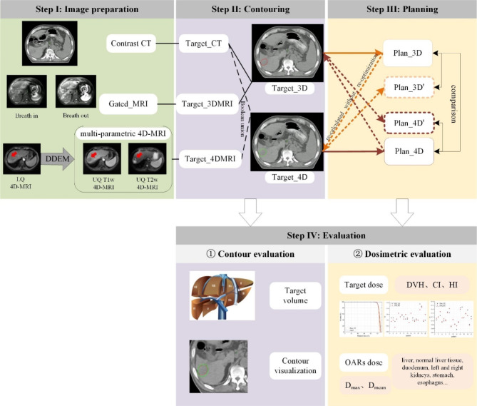
Methods: Twenty patients receiving radiotherapy for liver cancer were enrolled. Each patient underwent contrast-enhanced planning CT (free-breathing), contrast-enhanced T1-weighted (free-breathing), T2-weighted (gated) 3D-MRI, and low-quality 4D-MRI using the time resolved imaging with interleaved stochastic trajectories volumetric interpolated breath-hold examination (TWIST-VIBE) sequence. A dual-supervised deformation estimation model was used to generate a 4D deformable vector field (4D-DVF) from 4D-MRI data, and the prior images were deformed using this 4D-DVF to generate multi-parametric 4D-MRI. Assisted by 3D-MRI and multi-parametric 4D-MRI, target contours were performed on the planning CT, resulting in the generation of Target_3D and Target_4D. Clinical plans, Plan_3D and Plan_4D, were designed based on these contours respectively. To explore the dosimetric variations resulting from different contours without re-optimization, Plan_3D was directly applied to Target_4D, and Plan_4D was applied to Target_3D to generate Plan_3D' and Plan_4D' respectively. Target volume, contours, dose-volume histograms (DVHs), conformity index (CI), homogeneity index (HI), maximum and mean dose to organ as risks (OARs) were compared and evaluated.
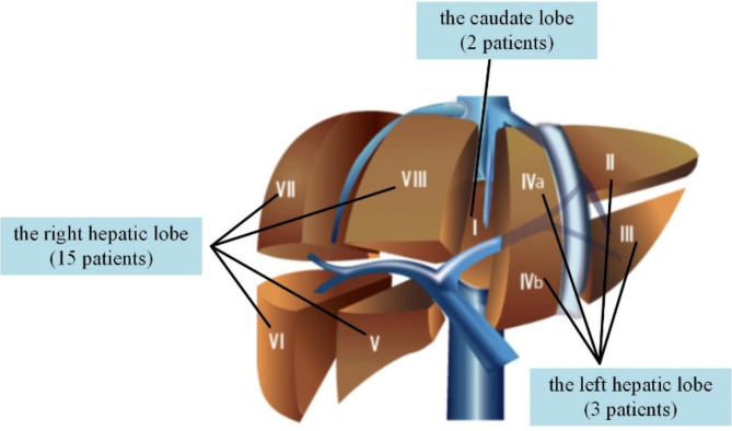
Results: Mean volume differences between Target_3D and Target_4D were 2.76 cm3 (standard deviation [SD] 3.42 cm3) in the caudate lobe, 181.54 cm3 (SD 68.50 cm3) in the left hepatic lobe, and 26.08 cm3 (SD 20.52 cm3) in the right hepatic lobe. Mean and SD of CI and HI is 1.02 ± 0.04 and 0.108 ± 0.02 in Plan_3D, 1.02 ± 0.01 and 0.107 ± 0.01 in Plan_4D. There were no statistically significant differences in OAR doses between Plan_3D and Plan_3D', between Plan_4D and Plan_4D'. However, a statistically significant difference in target dose was observed between Plan_3D and Plan_3D' (P = 1.47 × 10⁻⁷) and between Plan_4D and Plan_4D' (P = 0.013). Plan_3D' meets 100% of the prescription dose covering mean 77.89% (SD 10.13%) of the Targeted_4D volume, while Plan_4D' covered mean 94.17% (SD 3.12%) of the Targeted_3D volume.
Conclusions: 3D image-guided target delineation may be more likely to underestimate target volume and compromise dose coverage, suggesting that using multi-parametric 4D-MRI can provide more precise target contours and enhance target dose coverage.
Radiation OncologyONCOLOGY-RADIOLOGY, NUCLEAR MEDICINE & MEDICAL IMAGING
CiteScore
6.50
自引率
2.80%
发文量
181
审稿时长
3-6 weeks
期刊介绍:
Radiation Oncology encompasses all aspects of research that impacts on the treatment of cancer using radiation. It publishes findings in molecular and cellular radiation biology, radiation physics, radiation technology, and clinical oncology.
 求助内容:
求助内容: 应助结果提醒方式:


