Francesca Treballi, Ginevra Danti, Sofia Boccioli, Sebastiano Paolucci, Simone Busoni, Linda Calistri, Vittorio Miele
{"title":"直肠系膜脂肪的放射学特征作为直肠癌患者接受新辅助治疗的反应指标。","authors":"Francesca Treballi, Ginevra Danti, Sofia Boccioli, Sebastiano Paolucci, Simone Busoni, Linda Calistri, Vittorio Miele","doi":"10.3390/tomography11040044","DOIUrl":null,"url":null,"abstract":"<p><strong>Background: </strong>Rectal cancer represents a major cause of mortality in the United States. Management strategies are highly individualized, depending on patient-specific factors and tumor characteristics. The therapeutic landscape is rapidly evolving, with notable advancements in response rates to both radiotherapy and chemotherapy. For locally advanced rectal cancer (LARC, defined as up to T3-4 N+), the standard of care involves total mesorectal excision (TME) following neoadjuvant chemoradiotherapy (nCRT). Magnetic resonance imaging (MRI) has emerged as the gold standard for local tumor staging and is increasingly pivotal in post-treatment restaging.</p><p><strong>Aim: </strong>In our study, we proposed an MRI-based radiomic model to identify characteristic features of peritumoral mesorectal fat in two patient groups: good responders and poor responders to neoadjuvant therapy. The aim was to assess the potential presence of predictive factors for favorable or unfavorable responses to neoadjuvant chemoradiotherapy, thereby optimizing treatment management and improving personalized clinical decision-making.</p><p><strong>Methods: </strong>We conducted a retrospective analysis of adult patients with LARC who underwent pre- and post-nCRT MRI scans. Patients were classified as good responders (Group 0) or poor responders (Group 1) based on MRI findings, including tumor volume reduction, signal intensity changes on T2-weighted and diffusion-weighted imaging (DWI), and alterations in the circumferential resection margin (CRM) and extramural vascular invasion (EMVI) status. Classification criteria were based on the established literature to ensure consistency. Key clinical and imaging parameters, such as age, TNM stage, CRM involvement, and EMVI presence, were recorded. A radiomic model was developed using the LASSO algorithm for feature selection and regularization from 107 extracted radiomic features.</p><p><strong>Results: </strong>We included 44 patients (26 males and 18 females) who, following nCRT, were categorized into Group 0 (28 patients) and Group 1 (16 patients). The pre-treatment MRI analysis identified significant features (out of 107) for each sequence based on the Mann-Whitney test and <i>t</i>-test. The LASSO algorithm selected three features (shape_Sphericity, shape_Maximum2DDiameterSlice, and glcm_Imc2) for the construction of the radiomic logistic regression model, and ROC curves were subsequently generated for each model (AUC: 0.76).</p><p><strong>Conclusions: </strong>We developed an MRI-based radiomic model capable of differentiating and predicting between two groups of rectal cancer patients: responders and non-responders to neoadjuvant chemoradiotherapy (nCRT). This model has the potential to identify, at an early stage, lesions with a high likelihood of requiring surgery and those that could potentially be managed with medical treatment alone.</p>","PeriodicalId":51330,"journal":{"name":"Tomography","volume":"11 4","pages":""},"PeriodicalIF":2.2000,"publicationDate":"2025-04-07","publicationTypes":"Journal Article","fieldsOfStudy":null,"isOpenAccess":false,"openAccessPdf":"https://www.ncbi.nlm.nih.gov/pmc/articles/PMC12031397/pdf/","citationCount":"0","resultStr":"{\"title\":\"Radiomic Features of Mesorectal Fat as Indicators of Response in Rectal Cancer Patients Undergoing Neoadjuvant Therapy.\",\"authors\":\"Francesca Treballi, Ginevra Danti, Sofia Boccioli, Sebastiano Paolucci, Simone Busoni, Linda Calistri, Vittorio Miele\",\"doi\":\"10.3390/tomography11040044\",\"DOIUrl\":null,\"url\":null,\"abstract\":\"<p><strong>Background: </strong>Rectal cancer represents a major cause of mortality in the United States. Management strategies are highly individualized, depending on patient-specific factors and tumor characteristics. The therapeutic landscape is rapidly evolving, with notable advancements in response rates to both radiotherapy and chemotherapy. For locally advanced rectal cancer (LARC, defined as up to T3-4 N+), the standard of care involves total mesorectal excision (TME) following neoadjuvant chemoradiotherapy (nCRT). Magnetic resonance imaging (MRI) has emerged as the gold standard for local tumor staging and is increasingly pivotal in post-treatment restaging.</p><p><strong>Aim: </strong>In our study, we proposed an MRI-based radiomic model to identify characteristic features of peritumoral mesorectal fat in two patient groups: good responders and poor responders to neoadjuvant therapy. The aim was to assess the potential presence of predictive factors for favorable or unfavorable responses to neoadjuvant chemoradiotherapy, thereby optimizing treatment management and improving personalized clinical decision-making.</p><p><strong>Methods: </strong>We conducted a retrospective analysis of adult patients with LARC who underwent pre- and post-nCRT MRI scans. Patients were classified as good responders (Group 0) or poor responders (Group 1) based on MRI findings, including tumor volume reduction, signal intensity changes on T2-weighted and diffusion-weighted imaging (DWI), and alterations in the circumferential resection margin (CRM) and extramural vascular invasion (EMVI) status. Classification criteria were based on the established literature to ensure consistency. Key clinical and imaging parameters, such as age, TNM stage, CRM involvement, and EMVI presence, were recorded. A radiomic model was developed using the LASSO algorithm for feature selection and regularization from 107 extracted radiomic features.</p><p><strong>Results: </strong>We included 44 patients (26 males and 18 females) who, following nCRT, were categorized into Group 0 (28 patients) and Group 1 (16 patients). The pre-treatment MRI analysis identified significant features (out of 107) for each sequence based on the Mann-Whitney test and <i>t</i>-test. The LASSO algorithm selected three features (shape_Sphericity, shape_Maximum2DDiameterSlice, and glcm_Imc2) for the construction of the radiomic logistic regression model, and ROC curves were subsequently generated for each model (AUC: 0.76).</p><p><strong>Conclusions: </strong>We developed an MRI-based radiomic model capable of differentiating and predicting between two groups of rectal cancer patients: responders and non-responders to neoadjuvant chemoradiotherapy (nCRT). This model has the potential to identify, at an early stage, lesions with a high likelihood of requiring surgery and those that could potentially be managed with medical treatment alone.</p>\",\"PeriodicalId\":51330,\"journal\":{\"name\":\"Tomography\",\"volume\":\"11 4\",\"pages\":\"\"},\"PeriodicalIF\":2.2000,\"publicationDate\":\"2025-04-07\",\"publicationTypes\":\"Journal Article\",\"fieldsOfStudy\":null,\"isOpenAccess\":false,\"openAccessPdf\":\"https://www.ncbi.nlm.nih.gov/pmc/articles/PMC12031397/pdf/\",\"citationCount\":\"0\",\"resultStr\":null,\"platform\":\"Semanticscholar\",\"paperid\":null,\"PeriodicalName\":\"Tomography\",\"FirstCategoryId\":\"3\",\"ListUrlMain\":\"https://doi.org/10.3390/tomography11040044\",\"RegionNum\":4,\"RegionCategory\":\"医学\",\"ArticlePicture\":[],\"TitleCN\":null,\"AbstractTextCN\":null,\"PMCID\":null,\"EPubDate\":\"\",\"PubModel\":\"\",\"JCR\":\"Q2\",\"JCRName\":\"RADIOLOGY, NUCLEAR MEDICINE & MEDICAL IMAGING\",\"Score\":null,\"Total\":0}","platform":"Semanticscholar","paperid":null,"PeriodicalName":"Tomography","FirstCategoryId":"3","ListUrlMain":"https://doi.org/10.3390/tomography11040044","RegionNum":4,"RegionCategory":"医学","ArticlePicture":[],"TitleCN":null,"AbstractTextCN":null,"PMCID":null,"EPubDate":"","PubModel":"","JCR":"Q2","JCRName":"RADIOLOGY, NUCLEAR MEDICINE & MEDICAL IMAGING","Score":null,"Total":0}
Radiomic Features of Mesorectal Fat as Indicators of Response in Rectal Cancer Patients Undergoing Neoadjuvant Therapy.
Background: Rectal cancer represents a major cause of mortality in the United States. Management strategies are highly individualized, depending on patient-specific factors and tumor characteristics. The therapeutic landscape is rapidly evolving, with notable advancements in response rates to both radiotherapy and chemotherapy. For locally advanced rectal cancer (LARC, defined as up to T3-4 N+), the standard of care involves total mesorectal excision (TME) following neoadjuvant chemoradiotherapy (nCRT). Magnetic resonance imaging (MRI) has emerged as the gold standard for local tumor staging and is increasingly pivotal in post-treatment restaging.
Aim: In our study, we proposed an MRI-based radiomic model to identify characteristic features of peritumoral mesorectal fat in two patient groups: good responders and poor responders to neoadjuvant therapy. The aim was to assess the potential presence of predictive factors for favorable or unfavorable responses to neoadjuvant chemoradiotherapy, thereby optimizing treatment management and improving personalized clinical decision-making.
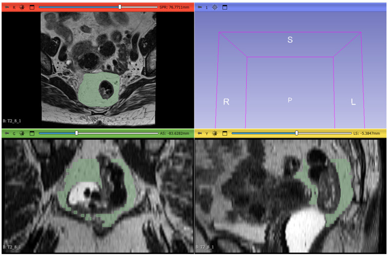
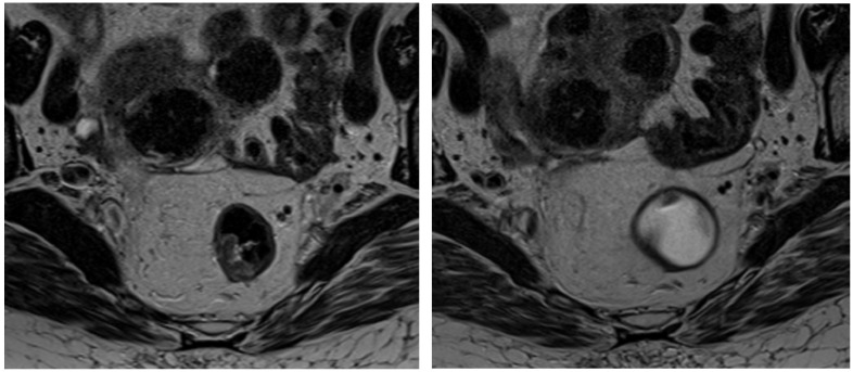
Methods: We conducted a retrospective analysis of adult patients with LARC who underwent pre- and post-nCRT MRI scans. Patients were classified as good responders (Group 0) or poor responders (Group 1) based on MRI findings, including tumor volume reduction, signal intensity changes on T2-weighted and diffusion-weighted imaging (DWI), and alterations in the circumferential resection margin (CRM) and extramural vascular invasion (EMVI) status. Classification criteria were based on the established literature to ensure consistency. Key clinical and imaging parameters, such as age, TNM stage, CRM involvement, and EMVI presence, were recorded. A radiomic model was developed using the LASSO algorithm for feature selection and regularization from 107 extracted radiomic features.
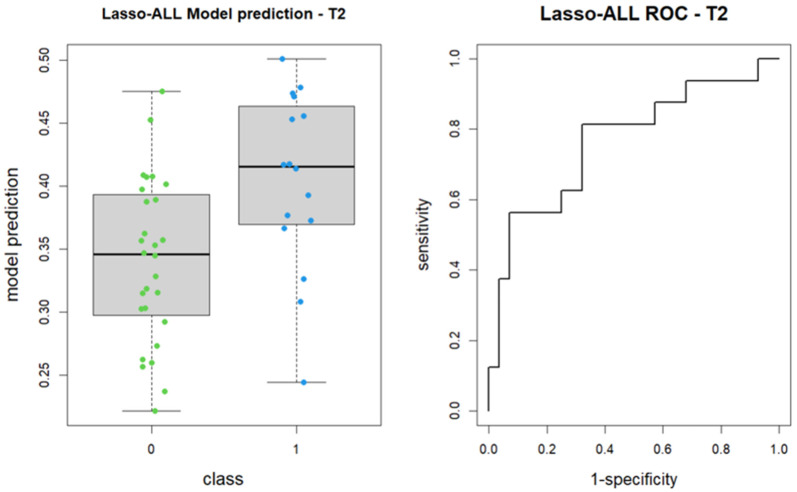
Results: We included 44 patients (26 males and 18 females) who, following nCRT, were categorized into Group 0 (28 patients) and Group 1 (16 patients). The pre-treatment MRI analysis identified significant features (out of 107) for each sequence based on the Mann-Whitney test and t-test. The LASSO algorithm selected three features (shape_Sphericity, shape_Maximum2DDiameterSlice, and glcm_Imc2) for the construction of the radiomic logistic regression model, and ROC curves were subsequently generated for each model (AUC: 0.76).
Conclusions: We developed an MRI-based radiomic model capable of differentiating and predicting between two groups of rectal cancer patients: responders and non-responders to neoadjuvant chemoradiotherapy (nCRT). This model has the potential to identify, at an early stage, lesions with a high likelihood of requiring surgery and those that could potentially be managed with medical treatment alone.
TomographyMedicine-Radiology, Nuclear Medicine and Imaging
CiteScore
2.70
自引率
10.50%
发文量
222
期刊介绍:
TomographyTM publishes basic (technical and pre-clinical) and clinical scientific articles which involve the advancement of imaging technologies. Tomography encompasses studies that use single or multiple imaging modalities including for example CT, US, PET, SPECT, MR and hyperpolarization technologies, as well as optical modalities (i.e. bioluminescence, photoacoustic, endomicroscopy, fiber optic imaging and optical computed tomography) in basic sciences, engineering, preclinical and clinical medicine.
Tomography also welcomes studies involving exploration and refinement of contrast mechanisms and image-derived metrics within and across modalities toward the development of novel imaging probes for image-based feedback and intervention. The use of imaging in biology and medicine provides unparalleled opportunities to noninvasively interrogate tissues to obtain real-time dynamic and quantitative information required for diagnosis and response to interventions and to follow evolving pathological conditions. As multi-modal studies and the complexities of imaging technologies themselves are ever increasing to provide advanced information to scientists and clinicians.
Tomography provides a unique publication venue allowing investigators the opportunity to more precisely communicate integrated findings related to the diverse and heterogeneous features associated with underlying anatomical, physiological, functional, metabolic and molecular genetic activities of normal and diseased tissue. Thus Tomography publishes peer-reviewed articles which involve the broad use of imaging of any tissue and disease type including both preclinical and clinical investigations. In addition, hardware/software along with chemical and molecular probe advances are welcome as they are deemed to significantly contribute towards the long-term goal of improving the overall impact of imaging on scientific and clinical discovery.

 求助内容:
求助内容: 应助结果提醒方式:


