{"title":"单侧海马硬化症颞叶癫痫患者的θ波振荡、颞顶和颞中枢网络的差异及其潜在机制。","authors":"Chenxi Qiu, Chenxi Zhong, Ying Liu, Liju Wang, Yingying Tang, Zhiyi Liu, Sijia Guo, Yingqi Jiang, Enzhi Li, Jing Lu, Bo Yan, Xiaoting Hao, Dong Zhou","doi":"10.1186/s42494-024-00170-7","DOIUrl":null,"url":null,"abstract":"<p><strong>Background: </strong>There is a lack of further exploration of the epileptogenic network of specific types of epilepsy, such as unilateral hippocampal sclerosis (HS), and there is an urgent need to find exact evidence to confirm the consistency of its brain network changes.</p><p><strong>Methods: </strong>We enrolled 22 mesial temporal lobe epilepsy with hippocampal sclerosis (mTLE-HS) patients to compare the differences in brain activity between 22 healthy controls (HCs) and them. Resting-state electroencephalography (EEG) was also measured. Then, we calculated the power spectral density and phase locking values in and between these electrodes.</p><p><strong>Results: </strong>The results showed the increased theta power was related to the high severity of epilepsy in the temporal, parietal, and central regions in mTLE-HS patients, and there were positive correlations between theta power in the contralateral temporal region and seizure frequency. Theta power in the ipsilateral parietal lobe is positively correlated with the number of anti-seizure medications (ASMs), but not with the usage of third-generation ASMs. Meanwhile, the temporal lobe of mTLE-HS patients had more connectivity with parietal lobe and central region.</p><p><strong>Conclusions: </strong>Theta power is an important EEG indicator of mTLE-HS, positively correlates with epilepsy severity and seizure frequency, and has network properties that can be observed outside the lesion. Moreover, the usage of third-generation ASMs did not affect the risk of increased theta power. Lastly, the temporoparietal and temporal-central networks are likely to be causative pathways in epilepsy patients with cognitive impairment. This study provides a potential guideline for the treatment of mTLE-HS in clinical practice.</p>","PeriodicalId":33628,"journal":{"name":"Acta Epileptologica","volume":"6 1","pages":"26"},"PeriodicalIF":1.2000,"publicationDate":"2024-08-25","publicationTypes":"Journal Article","fieldsOfStudy":null,"isOpenAccess":false,"openAccessPdf":"https://www.ncbi.nlm.nih.gov/pmc/articles/PMC11960361/pdf/","citationCount":"0","resultStr":"{\"title\":\"Differences and potential mechanisms of theta oscillation and temporoparietal and temporal-central networks in temporal lobe epilepsy patients with unilateral hippocampal sclerosis.\",\"authors\":\"Chenxi Qiu, Chenxi Zhong, Ying Liu, Liju Wang, Yingying Tang, Zhiyi Liu, Sijia Guo, Yingqi Jiang, Enzhi Li, Jing Lu, Bo Yan, Xiaoting Hao, Dong Zhou\",\"doi\":\"10.1186/s42494-024-00170-7\",\"DOIUrl\":null,\"url\":null,\"abstract\":\"<p><strong>Background: </strong>There is a lack of further exploration of the epileptogenic network of specific types of epilepsy, such as unilateral hippocampal sclerosis (HS), and there is an urgent need to find exact evidence to confirm the consistency of its brain network changes.</p><p><strong>Methods: </strong>We enrolled 22 mesial temporal lobe epilepsy with hippocampal sclerosis (mTLE-HS) patients to compare the differences in brain activity between 22 healthy controls (HCs) and them. Resting-state electroencephalography (EEG) was also measured. Then, we calculated the power spectral density and phase locking values in and between these electrodes.</p><p><strong>Results: </strong>The results showed the increased theta power was related to the high severity of epilepsy in the temporal, parietal, and central regions in mTLE-HS patients, and there were positive correlations between theta power in the contralateral temporal region and seizure frequency. Theta power in the ipsilateral parietal lobe is positively correlated with the number of anti-seizure medications (ASMs), but not with the usage of third-generation ASMs. Meanwhile, the temporal lobe of mTLE-HS patients had more connectivity with parietal lobe and central region.</p><p><strong>Conclusions: </strong>Theta power is an important EEG indicator of mTLE-HS, positively correlates with epilepsy severity and seizure frequency, and has network properties that can be observed outside the lesion. Moreover, the usage of third-generation ASMs did not affect the risk of increased theta power. Lastly, the temporoparietal and temporal-central networks are likely to be causative pathways in epilepsy patients with cognitive impairment. This study provides a potential guideline for the treatment of mTLE-HS in clinical practice.</p>\",\"PeriodicalId\":33628,\"journal\":{\"name\":\"Acta Epileptologica\",\"volume\":\"6 1\",\"pages\":\"26\"},\"PeriodicalIF\":1.2000,\"publicationDate\":\"2024-08-25\",\"publicationTypes\":\"Journal Article\",\"fieldsOfStudy\":null,\"isOpenAccess\":false,\"openAccessPdf\":\"https://www.ncbi.nlm.nih.gov/pmc/articles/PMC11960361/pdf/\",\"citationCount\":\"0\",\"resultStr\":null,\"platform\":\"Semanticscholar\",\"paperid\":null,\"PeriodicalName\":\"Acta Epileptologica\",\"FirstCategoryId\":\"1085\",\"ListUrlMain\":\"https://doi.org/10.1186/s42494-024-00170-7\",\"RegionNum\":0,\"RegionCategory\":null,\"ArticlePicture\":[],\"TitleCN\":null,\"AbstractTextCN\":null,\"PMCID\":null,\"EPubDate\":\"\",\"PubModel\":\"\",\"JCR\":\"Q4\",\"JCRName\":\"CLINICAL NEUROLOGY\",\"Score\":null,\"Total\":0}","platform":"Semanticscholar","paperid":null,"PeriodicalName":"Acta Epileptologica","FirstCategoryId":"1085","ListUrlMain":"https://doi.org/10.1186/s42494-024-00170-7","RegionNum":0,"RegionCategory":null,"ArticlePicture":[],"TitleCN":null,"AbstractTextCN":null,"PMCID":null,"EPubDate":"","PubModel":"","JCR":"Q4","JCRName":"CLINICAL NEUROLOGY","Score":null,"Total":0}
Differences and potential mechanisms of theta oscillation and temporoparietal and temporal-central networks in temporal lobe epilepsy patients with unilateral hippocampal sclerosis.
Background: There is a lack of further exploration of the epileptogenic network of specific types of epilepsy, such as unilateral hippocampal sclerosis (HS), and there is an urgent need to find exact evidence to confirm the consistency of its brain network changes.
Methods: We enrolled 22 mesial temporal lobe epilepsy with hippocampal sclerosis (mTLE-HS) patients to compare the differences in brain activity between 22 healthy controls (HCs) and them. Resting-state electroencephalography (EEG) was also measured. Then, we calculated the power spectral density and phase locking values in and between these electrodes.
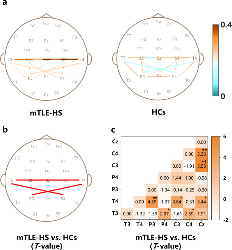
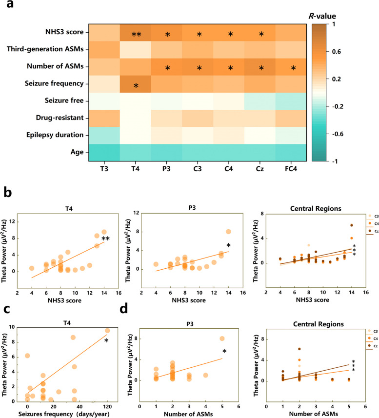
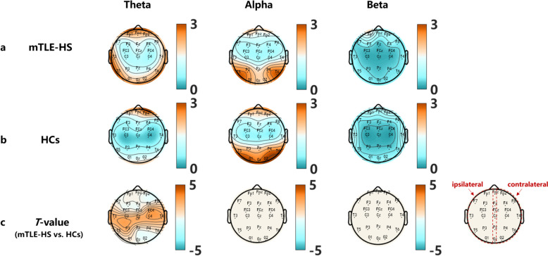
Results: The results showed the increased theta power was related to the high severity of epilepsy in the temporal, parietal, and central regions in mTLE-HS patients, and there were positive correlations between theta power in the contralateral temporal region and seizure frequency. Theta power in the ipsilateral parietal lobe is positively correlated with the number of anti-seizure medications (ASMs), but not with the usage of third-generation ASMs. Meanwhile, the temporal lobe of mTLE-HS patients had more connectivity with parietal lobe and central region.
Conclusions: Theta power is an important EEG indicator of mTLE-HS, positively correlates with epilepsy severity and seizure frequency, and has network properties that can be observed outside the lesion. Moreover, the usage of third-generation ASMs did not affect the risk of increased theta power. Lastly, the temporoparietal and temporal-central networks are likely to be causative pathways in epilepsy patients with cognitive impairment. This study provides a potential guideline for the treatment of mTLE-HS in clinical practice.
 求助内容:
求助内容: 应助结果提醒方式:


