Abdullah K Ghafour, Saywan K Asaad, Soran S Raoof, Rezheen J Rashid, Rebaz M Ali, Hiwa O Abdullah, Abdullah A Qadir, Shvan H Mohammed, Lawen Jamal Mustafa, Fahmi H Kakamad
{"title":"前臂近端不寻常的大骨旁脂肪瘤1例报告及文献复习。","authors":"Abdullah K Ghafour, Saywan K Asaad, Soran S Raoof, Rezheen J Rashid, Rebaz M Ali, Hiwa O Abdullah, Abdullah A Qadir, Shvan H Mohammed, Lawen Jamal Mustafa, Fahmi H Kakamad","doi":"10.1155/cro/7750483","DOIUrl":null,"url":null,"abstract":"<p><p><b>Introduction:</b> Parosteal lipomas are rare soft-tissue tumors with challenging surgical management. The current report is aimed at presenting a case of parosteal lipoma in a middle-aged female patient. <b>Case Presentation:</b> A 53-year-old diabetic lady presented with a gradually growing, painless mass in her right proximal forearm for the past 3 years. She complained of fatigue and reduced grip strength. The physical examination indicated a hard, immobile lump. A computed tomography scan revealed a clearly defined fat-density tumor with no bone involvement. Magnetic resonance imaging showed a well-defined fat-density mass surrounding most of the proximal radial shaft. The histological diagnosis of parosteal lipoma was made following surgical excision via Henry's approach. <b>Literature Review:</b> This minireview identified six reports on giant parosteal lipomas, involving patients aged adolescence to 83 years, mainly female. Common complaints were slowly progressive painless swellings. Imaging (radiographs, ultrasounds, CT, and MRI) revealed distinct features of lipomas. Surgical excision was the preferred management, with histopathology confirming lipoma diagnoses. Postoperative outcomes were positive, with no major complications or recurrences reported during follow-up. <b>Conclusion:</b> The tumor is a rare osseous neoplasm that may remain asymptomatic for years until it reaches a size capable of exerting pressure and causing motion difficulty. Meticulous care is paramount during surgical management to prevent iatrogenic nerve injury.</p>","PeriodicalId":30287,"journal":{"name":"Case Reports in Orthopedics","volume":"2025 ","pages":"7750483"},"PeriodicalIF":0.6000,"publicationDate":"2025-04-11","publicationTypes":"Journal Article","fieldsOfStudy":null,"isOpenAccess":false,"openAccessPdf":"https://www.ncbi.nlm.nih.gov/pmc/articles/PMC12008475/pdf/","citationCount":"0","resultStr":"{\"title\":\"Unusual Large Parosteal Lipoma of the Proximal Forearm: A Case Report and Literature Review.\",\"authors\":\"Abdullah K Ghafour, Saywan K Asaad, Soran S Raoof, Rezheen J Rashid, Rebaz M Ali, Hiwa O Abdullah, Abdullah A Qadir, Shvan H Mohammed, Lawen Jamal Mustafa, Fahmi H Kakamad\",\"doi\":\"10.1155/cro/7750483\",\"DOIUrl\":null,\"url\":null,\"abstract\":\"<p><p><b>Introduction:</b> Parosteal lipomas are rare soft-tissue tumors with challenging surgical management. The current report is aimed at presenting a case of parosteal lipoma in a middle-aged female patient. <b>Case Presentation:</b> A 53-year-old diabetic lady presented with a gradually growing, painless mass in her right proximal forearm for the past 3 years. She complained of fatigue and reduced grip strength. The physical examination indicated a hard, immobile lump. A computed tomography scan revealed a clearly defined fat-density tumor with no bone involvement. Magnetic resonance imaging showed a well-defined fat-density mass surrounding most of the proximal radial shaft. The histological diagnosis of parosteal lipoma was made following surgical excision via Henry's approach. <b>Literature Review:</b> This minireview identified six reports on giant parosteal lipomas, involving patients aged adolescence to 83 years, mainly female. Common complaints were slowly progressive painless swellings. Imaging (radiographs, ultrasounds, CT, and MRI) revealed distinct features of lipomas. Surgical excision was the preferred management, with histopathology confirming lipoma diagnoses. Postoperative outcomes were positive, with no major complications or recurrences reported during follow-up. <b>Conclusion:</b> The tumor is a rare osseous neoplasm that may remain asymptomatic for years until it reaches a size capable of exerting pressure and causing motion difficulty. Meticulous care is paramount during surgical management to prevent iatrogenic nerve injury.</p>\",\"PeriodicalId\":30287,\"journal\":{\"name\":\"Case Reports in Orthopedics\",\"volume\":\"2025 \",\"pages\":\"7750483\"},\"PeriodicalIF\":0.6000,\"publicationDate\":\"2025-04-11\",\"publicationTypes\":\"Journal Article\",\"fieldsOfStudy\":null,\"isOpenAccess\":false,\"openAccessPdf\":\"https://www.ncbi.nlm.nih.gov/pmc/articles/PMC12008475/pdf/\",\"citationCount\":\"0\",\"resultStr\":null,\"platform\":\"Semanticscholar\",\"paperid\":null,\"PeriodicalName\":\"Case Reports in Orthopedics\",\"FirstCategoryId\":\"1085\",\"ListUrlMain\":\"https://doi.org/10.1155/cro/7750483\",\"RegionNum\":0,\"RegionCategory\":null,\"ArticlePicture\":[],\"TitleCN\":null,\"AbstractTextCN\":null,\"PMCID\":null,\"EPubDate\":\"2025/1/1 0:00:00\",\"PubModel\":\"eCollection\",\"JCR\":\"Q4\",\"JCRName\":\"ORTHOPEDICS\",\"Score\":null,\"Total\":0}","platform":"Semanticscholar","paperid":null,"PeriodicalName":"Case Reports in Orthopedics","FirstCategoryId":"1085","ListUrlMain":"https://doi.org/10.1155/cro/7750483","RegionNum":0,"RegionCategory":null,"ArticlePicture":[],"TitleCN":null,"AbstractTextCN":null,"PMCID":null,"EPubDate":"2025/1/1 0:00:00","PubModel":"eCollection","JCR":"Q4","JCRName":"ORTHOPEDICS","Score":null,"Total":0}
Unusual Large Parosteal Lipoma of the Proximal Forearm: A Case Report and Literature Review.
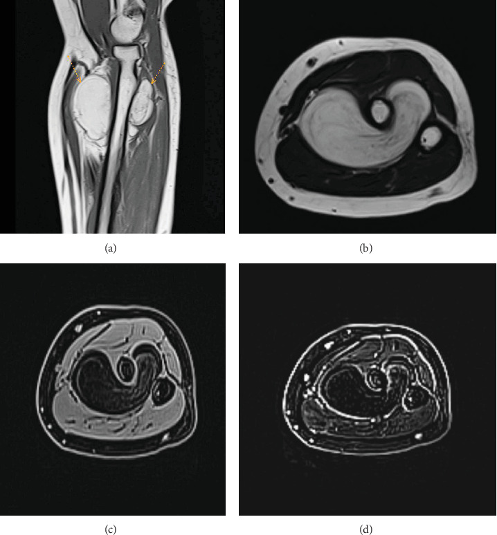
Introduction: Parosteal lipomas are rare soft-tissue tumors with challenging surgical management. The current report is aimed at presenting a case of parosteal lipoma in a middle-aged female patient. Case Presentation: A 53-year-old diabetic lady presented with a gradually growing, painless mass in her right proximal forearm for the past 3 years. She complained of fatigue and reduced grip strength. The physical examination indicated a hard, immobile lump. A computed tomography scan revealed a clearly defined fat-density tumor with no bone involvement. Magnetic resonance imaging showed a well-defined fat-density mass surrounding most of the proximal radial shaft. The histological diagnosis of parosteal lipoma was made following surgical excision via Henry's approach. Literature Review: This minireview identified six reports on giant parosteal lipomas, involving patients aged adolescence to 83 years, mainly female. Common complaints were slowly progressive painless swellings. Imaging (radiographs, ultrasounds, CT, and MRI) revealed distinct features of lipomas. Surgical excision was the preferred management, with histopathology confirming lipoma diagnoses. Postoperative outcomes were positive, with no major complications or recurrences reported during follow-up. Conclusion: The tumor is a rare osseous neoplasm that may remain asymptomatic for years until it reaches a size capable of exerting pressure and causing motion difficulty. Meticulous care is paramount during surgical management to prevent iatrogenic nerve injury.

 求助内容:
求助内容: 应助结果提醒方式:


