{"title":"从肩胛前上神经阻滞到膈神经的通路:尸体染色研究。","authors":"Sandeep Diwan, Deepika Sathe, Anjali Sabnis, Prakash Mane, Anju Gupta","doi":"10.17085/apm.24142","DOIUrl":null,"url":null,"abstract":"<p><strong>Background: </strong>Anterior suprascapular nerve block is widely used for postoperative shoulder pain management. Although cadaveric studies show the spread and smudging of the phrenic nerve, our cadaveric injection investigation was conducted to investigate the pathway of diffusion of the dye toward the phrenic nerve from a more distal injection at the suprascapular nerve.</p><p><strong>Methods: </strong>We injected 5 ml of 0.1% methylene blue dye into the proximal portion of the suprascapular nerve (infra-omohyoid in the posterior triangle) in 12 neck specimens from six cadavers. Following meticulous dissection, we assessed the spread of the dye along the brachial plexus to the nerve roots and traced the phrenic nerve for staining.</p><p><strong>Results: </strong>The phrenic nerve was stained in 41.7% of the cases, the inferior trunk of the brachial plexus was unstained in all cases (100%), and the posterior division and suprascapular nerve were stained in all cases (100%). The nerves to the subclavius, dorsal aspect of the superior trunk, and C5 and C6 roots were stained in all cases. Anterior division of the superior trunk was observed in 75% of the specimens. The dye-spread pathway along the brachial plexus was dorsal, sparing the ventral aspect.</p><p><strong>Conclusions: </strong>Our study demonstrated that the dye-spread pathway from the suprascapular nerve at the infra-omohyoid level to the phrenic nerve is dorsal to the brachial plexus.</p>","PeriodicalId":101360,"journal":{"name":"Anesthesia and pain medicine","volume":"20 2","pages":"175-182"},"PeriodicalIF":3.2000,"publicationDate":"2025-04-01","publicationTypes":"Journal Article","fieldsOfStudy":null,"isOpenAccess":false,"openAccessPdf":"https://www.ncbi.nlm.nih.gov/pmc/articles/PMC12066203/pdf/","citationCount":"0","resultStr":"{\"title\":\"Pathway from anterior suprascapular nerve block to the phrenic nerve: a cadaveric dye study.\",\"authors\":\"Sandeep Diwan, Deepika Sathe, Anjali Sabnis, Prakash Mane, Anju Gupta\",\"doi\":\"10.17085/apm.24142\",\"DOIUrl\":null,\"url\":null,\"abstract\":\"<p><strong>Background: </strong>Anterior suprascapular nerve block is widely used for postoperative shoulder pain management. Although cadaveric studies show the spread and smudging of the phrenic nerve, our cadaveric injection investigation was conducted to investigate the pathway of diffusion of the dye toward the phrenic nerve from a more distal injection at the suprascapular nerve.</p><p><strong>Methods: </strong>We injected 5 ml of 0.1% methylene blue dye into the proximal portion of the suprascapular nerve (infra-omohyoid in the posterior triangle) in 12 neck specimens from six cadavers. Following meticulous dissection, we assessed the spread of the dye along the brachial plexus to the nerve roots and traced the phrenic nerve for staining.</p><p><strong>Results: </strong>The phrenic nerve was stained in 41.7% of the cases, the inferior trunk of the brachial plexus was unstained in all cases (100%), and the posterior division and suprascapular nerve were stained in all cases (100%). The nerves to the subclavius, dorsal aspect of the superior trunk, and C5 and C6 roots were stained in all cases. Anterior division of the superior trunk was observed in 75% of the specimens. The dye-spread pathway along the brachial plexus was dorsal, sparing the ventral aspect.</p><p><strong>Conclusions: </strong>Our study demonstrated that the dye-spread pathway from the suprascapular nerve at the infra-omohyoid level to the phrenic nerve is dorsal to the brachial plexus.</p>\",\"PeriodicalId\":101360,\"journal\":{\"name\":\"Anesthesia and pain medicine\",\"volume\":\"20 2\",\"pages\":\"175-182\"},\"PeriodicalIF\":3.2000,\"publicationDate\":\"2025-04-01\",\"publicationTypes\":\"Journal Article\",\"fieldsOfStudy\":null,\"isOpenAccess\":false,\"openAccessPdf\":\"https://www.ncbi.nlm.nih.gov/pmc/articles/PMC12066203/pdf/\",\"citationCount\":\"0\",\"resultStr\":null,\"platform\":\"Semanticscholar\",\"paperid\":null,\"PeriodicalName\":\"Anesthesia and pain medicine\",\"FirstCategoryId\":\"1085\",\"ListUrlMain\":\"https://doi.org/10.17085/apm.24142\",\"RegionNum\":0,\"RegionCategory\":null,\"ArticlePicture\":[],\"TitleCN\":null,\"AbstractTextCN\":null,\"PMCID\":null,\"EPubDate\":\"2025/4/30 0:00:00\",\"PubModel\":\"Epub\",\"JCR\":\"\",\"JCRName\":\"\",\"Score\":null,\"Total\":0}","platform":"Semanticscholar","paperid":null,"PeriodicalName":"Anesthesia and pain medicine","FirstCategoryId":"1085","ListUrlMain":"https://doi.org/10.17085/apm.24142","RegionNum":0,"RegionCategory":null,"ArticlePicture":[],"TitleCN":null,"AbstractTextCN":null,"PMCID":null,"EPubDate":"2025/4/30 0:00:00","PubModel":"Epub","JCR":"","JCRName":"","Score":null,"Total":0}
Pathway from anterior suprascapular nerve block to the phrenic nerve: a cadaveric dye study.
Background: Anterior suprascapular nerve block is widely used for postoperative shoulder pain management. Although cadaveric studies show the spread and smudging of the phrenic nerve, our cadaveric injection investigation was conducted to investigate the pathway of diffusion of the dye toward the phrenic nerve from a more distal injection at the suprascapular nerve.
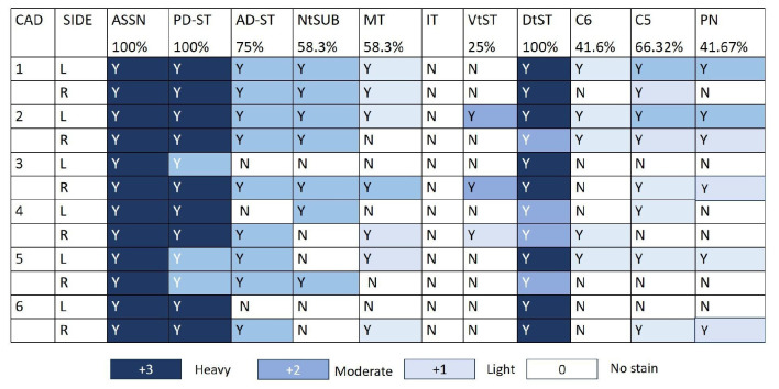
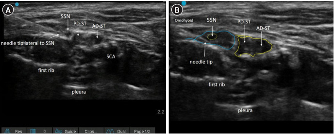
Methods: We injected 5 ml of 0.1% methylene blue dye into the proximal portion of the suprascapular nerve (infra-omohyoid in the posterior triangle) in 12 neck specimens from six cadavers. Following meticulous dissection, we assessed the spread of the dye along the brachial plexus to the nerve roots and traced the phrenic nerve for staining.
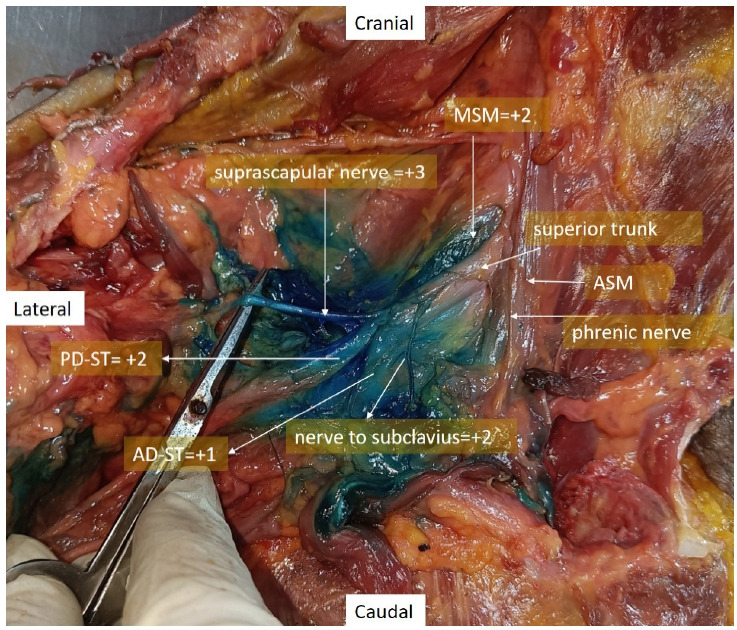
Results: The phrenic nerve was stained in 41.7% of the cases, the inferior trunk of the brachial plexus was unstained in all cases (100%), and the posterior division and suprascapular nerve were stained in all cases (100%). The nerves to the subclavius, dorsal aspect of the superior trunk, and C5 and C6 roots were stained in all cases. Anterior division of the superior trunk was observed in 75% of the specimens. The dye-spread pathway along the brachial plexus was dorsal, sparing the ventral aspect.
Conclusions: Our study demonstrated that the dye-spread pathway from the suprascapular nerve at the infra-omohyoid level to the phrenic nerve is dorsal to the brachial plexus.

 求助内容:
求助内容: 应助结果提醒方式:


