{"title":"隐藏在普通视野:未知解剖描述和应用的主动脉-食管韧带。","authors":"Nanditha Guruvaiah Sridhara, Namratha Guruvaiah Sridhara, Janardhana Ponnatapura","doi":"10.21037/med-24-31","DOIUrl":null,"url":null,"abstract":"<p><p>While it is not uncommon to see central mediastinal diseases on cross-sectional imaging, it is important to understand the pathway influencing the spread of disease at a radiological point of view. The advent of minimally invasive thoracic surgeries has led to the discovery of unknown tissue planes in the mediastinum such as the aorto-esophageal (AE) and aorto-pleural (AP) ligaments. In particular, the AE ligament is a portion of the mediastinal visceral fascia, which courses from the anterior aspect of the aorta to the left lateral aspect of the esophagus. It can be visualized on computed tomography (CT) and magnetic resonance imaging (MRI); it courses longitudinally from the level of the aortic arch to the level of the diaphragm. This recently discovered unknown anatomy aids us in understanding the possible pathway of spread of disease processes such as air, fluid, and soft tissue in the mediastinum. In addition, it acts as an important anatomical landmark in determining the location of lymph node metastases from esophageal cancer, which will further influence the possibility of thoracic duct resection/sparing. Finally, the AE ligament can be utilized in the preoperative planning of minimally invasive thoracic surgeries and can potentially be used as a dissection plane during esophagectomies.</p>","PeriodicalId":74139,"journal":{"name":"Mediastinum (Hong Kong, China)","volume":"9 ","pages":"3"},"PeriodicalIF":0.0000,"publicationDate":"2025-03-06","publicationTypes":"Journal Article","fieldsOfStudy":null,"isOpenAccess":false,"openAccessPdf":"https://www.ncbi.nlm.nih.gov/pmc/articles/PMC11982987/pdf/","citationCount":"0","resultStr":"{\"title\":\"Hidden in plain sight: unknown anatomy depiction and applications of the aorto-esophageal ligament.\",\"authors\":\"Nanditha Guruvaiah Sridhara, Namratha Guruvaiah Sridhara, Janardhana Ponnatapura\",\"doi\":\"10.21037/med-24-31\",\"DOIUrl\":null,\"url\":null,\"abstract\":\"<p><p>While it is not uncommon to see central mediastinal diseases on cross-sectional imaging, it is important to understand the pathway influencing the spread of disease at a radiological point of view. The advent of minimally invasive thoracic surgeries has led to the discovery of unknown tissue planes in the mediastinum such as the aorto-esophageal (AE) and aorto-pleural (AP) ligaments. In particular, the AE ligament is a portion of the mediastinal visceral fascia, which courses from the anterior aspect of the aorta to the left lateral aspect of the esophagus. It can be visualized on computed tomography (CT) and magnetic resonance imaging (MRI); it courses longitudinally from the level of the aortic arch to the level of the diaphragm. This recently discovered unknown anatomy aids us in understanding the possible pathway of spread of disease processes such as air, fluid, and soft tissue in the mediastinum. In addition, it acts as an important anatomical landmark in determining the location of lymph node metastases from esophageal cancer, which will further influence the possibility of thoracic duct resection/sparing. Finally, the AE ligament can be utilized in the preoperative planning of minimally invasive thoracic surgeries and can potentially be used as a dissection plane during esophagectomies.</p>\",\"PeriodicalId\":74139,\"journal\":{\"name\":\"Mediastinum (Hong Kong, China)\",\"volume\":\"9 \",\"pages\":\"3\"},\"PeriodicalIF\":0.0000,\"publicationDate\":\"2025-03-06\",\"publicationTypes\":\"Journal Article\",\"fieldsOfStudy\":null,\"isOpenAccess\":false,\"openAccessPdf\":\"https://www.ncbi.nlm.nih.gov/pmc/articles/PMC11982987/pdf/\",\"citationCount\":\"0\",\"resultStr\":null,\"platform\":\"Semanticscholar\",\"paperid\":null,\"PeriodicalName\":\"Mediastinum (Hong Kong, China)\",\"FirstCategoryId\":\"1085\",\"ListUrlMain\":\"https://doi.org/10.21037/med-24-31\",\"RegionNum\":0,\"RegionCategory\":null,\"ArticlePicture\":[],\"TitleCN\":null,\"AbstractTextCN\":null,\"PMCID\":null,\"EPubDate\":\"2025/1/1 0:00:00\",\"PubModel\":\"eCollection\",\"JCR\":\"\",\"JCRName\":\"\",\"Score\":null,\"Total\":0}","platform":"Semanticscholar","paperid":null,"PeriodicalName":"Mediastinum (Hong Kong, China)","FirstCategoryId":"1085","ListUrlMain":"https://doi.org/10.21037/med-24-31","RegionNum":0,"RegionCategory":null,"ArticlePicture":[],"TitleCN":null,"AbstractTextCN":null,"PMCID":null,"EPubDate":"2025/1/1 0:00:00","PubModel":"eCollection","JCR":"","JCRName":"","Score":null,"Total":0}
Hidden in plain sight: unknown anatomy depiction and applications of the aorto-esophageal ligament.
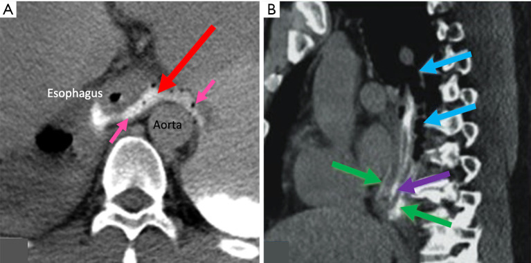
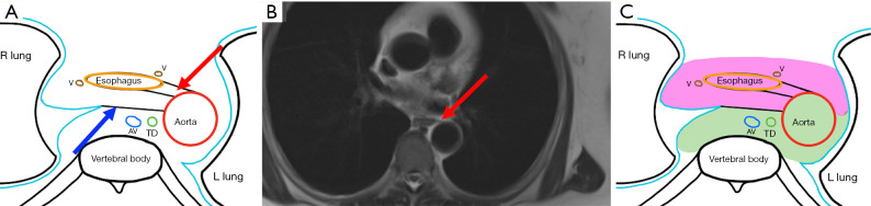
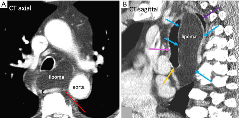
While it is not uncommon to see central mediastinal diseases on cross-sectional imaging, it is important to understand the pathway influencing the spread of disease at a radiological point of view. The advent of minimally invasive thoracic surgeries has led to the discovery of unknown tissue planes in the mediastinum such as the aorto-esophageal (AE) and aorto-pleural (AP) ligaments. In particular, the AE ligament is a portion of the mediastinal visceral fascia, which courses from the anterior aspect of the aorta to the left lateral aspect of the esophagus. It can be visualized on computed tomography (CT) and magnetic resonance imaging (MRI); it courses longitudinally from the level of the aortic arch to the level of the diaphragm. This recently discovered unknown anatomy aids us in understanding the possible pathway of spread of disease processes such as air, fluid, and soft tissue in the mediastinum. In addition, it acts as an important anatomical landmark in determining the location of lymph node metastases from esophageal cancer, which will further influence the possibility of thoracic duct resection/sparing. Finally, the AE ligament can be utilized in the preoperative planning of minimally invasive thoracic surgeries and can potentially be used as a dissection plane during esophagectomies.

 求助内容:
求助内容: 应助结果提醒方式:


