Beste M Atasoy, Birsen Demirel, Feyza Nur Ekşi Özdaş, Bennur Devran, Zehra Nur Kılıç, Dilek Gül
{"title":"放疗计划图像在监测头颈癌患者营养不良和预测预后中的作用:一项初步研究。","authors":"Beste M Atasoy, Birsen Demirel, Feyza Nur Ekşi Özdaş, Bennur Devran, Zehra Nur Kılıç, Dilek Gül","doi":"10.1186/s13014-025-02645-4","DOIUrl":null,"url":null,"abstract":"<p><strong>Background: </strong>Adaptive treatment planning can be made in radiotherapy of head and neck cancer patients for reasons such as changes in tumor volume or weight loss. This study aims to find the role of treatment planning images in monitoring radiotherapy-induced malnutrition and predicting the malnutrition-induced prognosis in head and neck cancer patients.</p><p><strong>Methods: </strong>For this study, we analyzed 30 patients who received radiotherapy in our clinic between September 2018 and September 2021. Those patients, both regular and completed weekly dietitian counseling notes during radiotherapy and available adaptive radiotherapy planning images, were included in the analysis. All patients had weekly nutritional interventions, including nutritional and anthropometric changes in weight, height, body mass index (BMI), and lean body mass (LBM). Skeletal muscle volume, called cervical muscle gauge (CMG), was measured from the simulation images of beginning and adaptive radiotherapy. Inflammatory parameters, including the neutrophil-lymphocyte ratio (NLR), the platelet-lymphocyte ratio (PLR), and the systemic inflammatory index (SII), were also calculated from weekly total blood counts. For the analysis, anthropometric measurements were compared at the beginning and adaptive treatment time. Progression-free (PFS) and overall (OS) survival were calculated according to weight and CMG changes.</p><p><strong>Results: </strong>The median weight loss percentage was 4.8% (0 to 24%). The mean percentage of weight changes, LBM, and CMG were 6.33%, 3.47%, and 9.28%, respectively. Results indicated that BMI (p = 006), weight (p < 0.001), LBM (p < 0.001), and CMG (p = 0.057) decreased during radiotherapy. Hemoglobin levels decreased (p = 0.005), and inflammatory markers increased. There were significant correlations between weight and LBM (p < 0.0001) and CMG (p = 0.005) loss. The median follow-up was 26 months. Loss of weight (PFS; 65.5% vs. 35.7%, p = 0.09, OS; 73.7% vs. 32.1%, p = 0.09), LBM (PFS; 75% vs. 41.1%, p = 0.118, OS; 65.6% vs. 52%, p = 0.221) and CMG (PFS; 56.3% vs. 47.1%, p = 0.516, OS;76.9% vs. 32.4%, p = 0.059) negatively affected three-year survival.</p><p><strong>Conclusions: </strong>Cervical muscle volume measurement may help predict malnutrition in patients receiving radiotherapy for head and neck cancer. Our study shows adaptive planning images may be used for this approach. In addition, this method may help to predict prognosis due to malnutrition in patients undergoing radiotherapy.</p>","PeriodicalId":49639,"journal":{"name":"Radiation Oncology","volume":"20 1","pages":"70"},"PeriodicalIF":3.3000,"publicationDate":"2025-05-03","publicationTypes":"Journal Article","fieldsOfStudy":null,"isOpenAccess":false,"openAccessPdf":"https://www.ncbi.nlm.nih.gov/pmc/articles/PMC12049786/pdf/","citationCount":"0","resultStr":"{\"title\":\"The role of radiotherapy planning images in monitoring malnutrition and predicting prognosis in head and neck cancer patients: a pilot study.\",\"authors\":\"Beste M Atasoy, Birsen Demirel, Feyza Nur Ekşi Özdaş, Bennur Devran, Zehra Nur Kılıç, Dilek Gül\",\"doi\":\"10.1186/s13014-025-02645-4\",\"DOIUrl\":null,\"url\":null,\"abstract\":\"<p><strong>Background: </strong>Adaptive treatment planning can be made in radiotherapy of head and neck cancer patients for reasons such as changes in tumor volume or weight loss. This study aims to find the role of treatment planning images in monitoring radiotherapy-induced malnutrition and predicting the malnutrition-induced prognosis in head and neck cancer patients.</p><p><strong>Methods: </strong>For this study, we analyzed 30 patients who received radiotherapy in our clinic between September 2018 and September 2021. Those patients, both regular and completed weekly dietitian counseling notes during radiotherapy and available adaptive radiotherapy planning images, were included in the analysis. All patients had weekly nutritional interventions, including nutritional and anthropometric changes in weight, height, body mass index (BMI), and lean body mass (LBM). Skeletal muscle volume, called cervical muscle gauge (CMG), was measured from the simulation images of beginning and adaptive radiotherapy. Inflammatory parameters, including the neutrophil-lymphocyte ratio (NLR), the platelet-lymphocyte ratio (PLR), and the systemic inflammatory index (SII), were also calculated from weekly total blood counts. For the analysis, anthropometric measurements were compared at the beginning and adaptive treatment time. Progression-free (PFS) and overall (OS) survival were calculated according to weight and CMG changes.</p><p><strong>Results: </strong>The median weight loss percentage was 4.8% (0 to 24%). The mean percentage of weight changes, LBM, and CMG were 6.33%, 3.47%, and 9.28%, respectively. Results indicated that BMI (p = 006), weight (p < 0.001), LBM (p < 0.001), and CMG (p = 0.057) decreased during radiotherapy. Hemoglobin levels decreased (p = 0.005), and inflammatory markers increased. There were significant correlations between weight and LBM (p < 0.0001) and CMG (p = 0.005) loss. The median follow-up was 26 months. Loss of weight (PFS; 65.5% vs. 35.7%, p = 0.09, OS; 73.7% vs. 32.1%, p = 0.09), LBM (PFS; 75% vs. 41.1%, p = 0.118, OS; 65.6% vs. 52%, p = 0.221) and CMG (PFS; 56.3% vs. 47.1%, p = 0.516, OS;76.9% vs. 32.4%, p = 0.059) negatively affected three-year survival.</p><p><strong>Conclusions: </strong>Cervical muscle volume measurement may help predict malnutrition in patients receiving radiotherapy for head and neck cancer. Our study shows adaptive planning images may be used for this approach. In addition, this method may help to predict prognosis due to malnutrition in patients undergoing radiotherapy.</p>\",\"PeriodicalId\":49639,\"journal\":{\"name\":\"Radiation Oncology\",\"volume\":\"20 1\",\"pages\":\"70\"},\"PeriodicalIF\":3.3000,\"publicationDate\":\"2025-05-03\",\"publicationTypes\":\"Journal Article\",\"fieldsOfStudy\":null,\"isOpenAccess\":false,\"openAccessPdf\":\"https://www.ncbi.nlm.nih.gov/pmc/articles/PMC12049786/pdf/\",\"citationCount\":\"0\",\"resultStr\":null,\"platform\":\"Semanticscholar\",\"paperid\":null,\"PeriodicalName\":\"Radiation Oncology\",\"FirstCategoryId\":\"3\",\"ListUrlMain\":\"https://doi.org/10.1186/s13014-025-02645-4\",\"RegionNum\":2,\"RegionCategory\":\"医学\",\"ArticlePicture\":[],\"TitleCN\":null,\"AbstractTextCN\":null,\"PMCID\":null,\"EPubDate\":\"\",\"PubModel\":\"\",\"JCR\":\"Q2\",\"JCRName\":\"ONCOLOGY\",\"Score\":null,\"Total\":0}","platform":"Semanticscholar","paperid":null,"PeriodicalName":"Radiation Oncology","FirstCategoryId":"3","ListUrlMain":"https://doi.org/10.1186/s13014-025-02645-4","RegionNum":2,"RegionCategory":"医学","ArticlePicture":[],"TitleCN":null,"AbstractTextCN":null,"PMCID":null,"EPubDate":"","PubModel":"","JCR":"Q2","JCRName":"ONCOLOGY","Score":null,"Total":0}
The role of radiotherapy planning images in monitoring malnutrition and predicting prognosis in head and neck cancer patients: a pilot study.
Background: Adaptive treatment planning can be made in radiotherapy of head and neck cancer patients for reasons such as changes in tumor volume or weight loss. This study aims to find the role of treatment planning images in monitoring radiotherapy-induced malnutrition and predicting the malnutrition-induced prognosis in head and neck cancer patients.
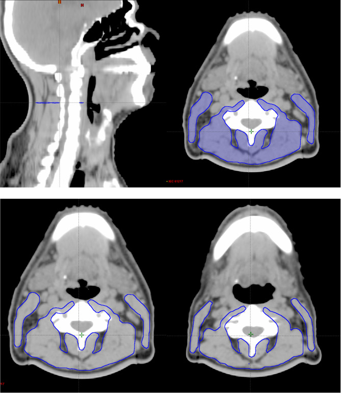
Methods: For this study, we analyzed 30 patients who received radiotherapy in our clinic between September 2018 and September 2021. Those patients, both regular and completed weekly dietitian counseling notes during radiotherapy and available adaptive radiotherapy planning images, were included in the analysis. All patients had weekly nutritional interventions, including nutritional and anthropometric changes in weight, height, body mass index (BMI), and lean body mass (LBM). Skeletal muscle volume, called cervical muscle gauge (CMG), was measured from the simulation images of beginning and adaptive radiotherapy. Inflammatory parameters, including the neutrophil-lymphocyte ratio (NLR), the platelet-lymphocyte ratio (PLR), and the systemic inflammatory index (SII), were also calculated from weekly total blood counts. For the analysis, anthropometric measurements were compared at the beginning and adaptive treatment time. Progression-free (PFS) and overall (OS) survival were calculated according to weight and CMG changes.
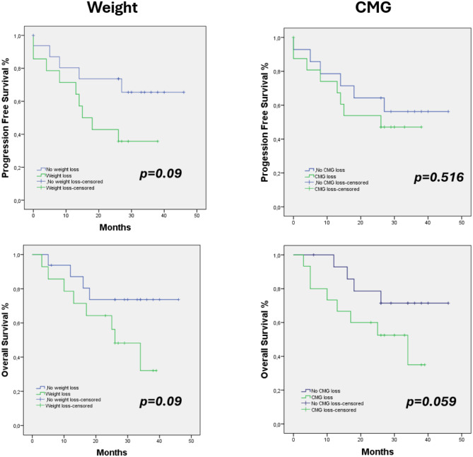
Results: The median weight loss percentage was 4.8% (0 to 24%). The mean percentage of weight changes, LBM, and CMG were 6.33%, 3.47%, and 9.28%, respectively. Results indicated that BMI (p = 006), weight (p < 0.001), LBM (p < 0.001), and CMG (p = 0.057) decreased during radiotherapy. Hemoglobin levels decreased (p = 0.005), and inflammatory markers increased. There were significant correlations between weight and LBM (p < 0.0001) and CMG (p = 0.005) loss. The median follow-up was 26 months. Loss of weight (PFS; 65.5% vs. 35.7%, p = 0.09, OS; 73.7% vs. 32.1%, p = 0.09), LBM (PFS; 75% vs. 41.1%, p = 0.118, OS; 65.6% vs. 52%, p = 0.221) and CMG (PFS; 56.3% vs. 47.1%, p = 0.516, OS;76.9% vs. 32.4%, p = 0.059) negatively affected three-year survival.
Conclusions: Cervical muscle volume measurement may help predict malnutrition in patients receiving radiotherapy for head and neck cancer. Our study shows adaptive planning images may be used for this approach. In addition, this method may help to predict prognosis due to malnutrition in patients undergoing radiotherapy.
Radiation OncologyONCOLOGY-RADIOLOGY, NUCLEAR MEDICINE & MEDICAL IMAGING
CiteScore
6.50
自引率
2.80%
发文量
181
审稿时长
3-6 weeks
期刊介绍:
Radiation Oncology encompasses all aspects of research that impacts on the treatment of cancer using radiation. It publishes findings in molecular and cellular radiation biology, radiation physics, radiation technology, and clinical oncology.
 求助内容:
求助内容: 应助结果提醒方式:


