评估妊娠中期先兆子痫的风险以确定后续护理。

IF 6.1 1区 医学 Q1 ACOUSTICS
Ultrasound in Obstetrics & Gynecology Pub Date : 2025-06-01 Epub Date: 2025-04-18 DOI:10.1002/uog.29222
S Adjahou, V Logdanidis, A Wright, A Syngelaki, R Akolekar, K H Nicolaides
{"title":"评估妊娠中期先兆子痫的风险以确定后续护理。","authors":"S Adjahou, V Logdanidis, A Wright, A Syngelaki, R Akolekar, K H Nicolaides","doi":"10.1002/uog.29222","DOIUrl":null,"url":null,"abstract":"<p><strong>Objective: </strong>To stratify pregnancy care based on the estimated risk of pre-eclampsia (PE) from screening at 19-24 weeks' gestation by combinations of maternal risk factors, estimated fetal weight (EFW), mean arterial pressure (MAP) and uterine artery pulsatility index (UtA-PI).</p><p><strong>Methods: </strong>The data for this study were derived from a prospective non-interventional study in 134 443 women with a singleton pregnancy attending for a routine ultrasound scan at 19 + 0 to 23 + 6 weeks' gestation in two UK maternity hospitals. The visit included recording of maternal demographic characteristics and medical history, sonographic EFW and measurement of MAP and UtA-PI. The competing-risks model was used to estimate the individual patient-specific risk of delivery with PE at < 28, < 32 and < 36 weeks' gestation. Receiver-operating-characteristics curves were constructed for screen-positive rates (SPRs) at different detection rates of delivery with PE at < 28, < 32 and < 36 weeks' gestation for the combinations of maternal risk factors, EFW and MAP, and of maternal risk factors, EFW, MAP and UtA-PI. Different risk cut-offs were used with the intention of detecting about 80%, 85% and 90% of cases of delivery with PE at < 28, < 32 and < 36 weeks' gestation. Calibration for risk of delivery with PE at < 28, < 32 and < 36 weeks' gestation was assessed by plotting the observed incidence of PE against the predicted incidence of PE.</p><p><strong>Results: </strong>The study population contained 4335 (3.2%) women that subsequently developed PE, including 64 (0.05%) that delivered with PE at < 28 weeks' gestation, 209 (0.2%) that delivered with PE at < 32 weeks and 655 (0.5%) that delivered with PE at < 36 weeks. If the objective of screening was to identify about 90% of cases of delivery with PE at < 28, < 32 and < 36 weeks and the method of screening was a combination of maternal risk factors, EFW and MAP, the respective SPRs would be 11.0%, 18.3% and 38.8%. If the method of screening also included UtA-PI, the respective SPRs would be 2.6%, 3.8% and 23.6%. If the objective of screening was to identify about 80% of cases of delivery with PE at < 28, < 32 and < 36 weeks and the method of screening was a combination of maternal risk factors, EFW and MAP, the respective SPRs would be 5.9%, 9.7% and 21.9%. If the method of screening also included UtA-PI, the respective SPRs would be 1.0%, 2.1% and 11.7%. The calibration plots demonstrated good agreement between the estimated risk and observed incidence of PE.</p><p><strong>Conclusions: </strong>All women should be offered assessment of risk for PE at 11-13 weeks, to help identify those requiring aspirin prophylaxis to reduce the rate of preterm PE, and at 35-37 weeks, to determine the optimal timing of birth to reduce the rate of term PE. Assessment of risk for PE at mid-gestation can be used to identify the subgroups that require additional monitoring at 24-35, 28-35 and 32-35 weeks' gestation. The best performance of screening, reflected in the SPR necessary to achieve a high detection rate, is achieved by a combination of maternal risk factors, MAP and UtA-PI. © 2025 The Author(s). Ultrasound in Obstetrics & Gynecology published by John Wiley & Sons Ltd on behalf of International Society of Ultrasound in Obstetrics and Gynecology.</p>","PeriodicalId":23454,"journal":{"name":"Ultrasound in Obstetrics & Gynecology","volume":" ","pages":"694-702"},"PeriodicalIF":6.1000,"publicationDate":"2025-06-01","publicationTypes":"Journal Article","fieldsOfStudy":null,"isOpenAccess":false,"openAccessPdf":"https://www.ncbi.nlm.nih.gov/pmc/articles/PMC12127725/pdf/","citationCount":"0","resultStr":"{\"title\":\"Assessment of risk for pre-eclampsia at mid-gestation to define subsequent care.\",\"authors\":\"S Adjahou, V Logdanidis, A Wright, A Syngelaki, R Akolekar, K H Nicolaides\",\"doi\":\"10.1002/uog.29222\",\"DOIUrl\":null,\"url\":null,\"abstract\":\"<p><strong>Objective: </strong>To stratify pregnancy care based on the estimated risk of pre-eclampsia (PE) from screening at 19-24 weeks' gestation by combinations of maternal risk factors, estimated fetal weight (EFW), mean arterial pressure (MAP) and uterine artery pulsatility index (UtA-PI).</p><p><strong>Methods: </strong>The data for this study were derived from a prospective non-interventional study in 134 443 women with a singleton pregnancy attending for a routine ultrasound scan at 19 + 0 to 23 + 6 weeks' gestation in two UK maternity hospitals. The visit included recording of maternal demographic characteristics and medical history, sonographic EFW and measurement of MAP and UtA-PI. The competing-risks model was used to estimate the individual patient-specific risk of delivery with PE at < 28, < 32 and < 36 weeks' gestation. Receiver-operating-characteristics curves were constructed for screen-positive rates (SPRs) at different detection rates of delivery with PE at < 28, < 32 and < 36 weeks' gestation for the combinations of maternal risk factors, EFW and MAP, and of maternal risk factors, EFW, MAP and UtA-PI. Different risk cut-offs were used with the intention of detecting about 80%, 85% and 90% of cases of delivery with PE at < 28, < 32 and < 36 weeks' gestation. Calibration for risk of delivery with PE at < 28, < 32 and < 36 weeks' gestation was assessed by plotting the observed incidence of PE against the predicted incidence of PE.</p><p><strong>Results: </strong>The study population contained 4335 (3.2%) women that subsequently developed PE, including 64 (0.05%) that delivered with PE at < 28 weeks' gestation, 209 (0.2%) that delivered with PE at < 32 weeks and 655 (0.5%) that delivered with PE at < 36 weeks. If the objective of screening was to identify about 90% of cases of delivery with PE at < 28, < 32 and < 36 weeks and the method of screening was a combination of maternal risk factors, EFW and MAP, the respective SPRs would be 11.0%, 18.3% and 38.8%. If the method of screening also included UtA-PI, the respective SPRs would be 2.6%, 3.8% and 23.6%. If the objective of screening was to identify about 80% of cases of delivery with PE at < 28, < 32 and < 36 weeks and the method of screening was a combination of maternal risk factors, EFW and MAP, the respective SPRs would be 5.9%, 9.7% and 21.9%. If the method of screening also included UtA-PI, the respective SPRs would be 1.0%, 2.1% and 11.7%. The calibration plots demonstrated good agreement between the estimated risk and observed incidence of PE.</p><p><strong>Conclusions: </strong>All women should be offered assessment of risk for PE at 11-13 weeks, to help identify those requiring aspirin prophylaxis to reduce the rate of preterm PE, and at 35-37 weeks, to determine the optimal timing of birth to reduce the rate of term PE. Assessment of risk for PE at mid-gestation can be used to identify the subgroups that require additional monitoring at 24-35, 28-35 and 32-35 weeks' gestation. The best performance of screening, reflected in the SPR necessary to achieve a high detection rate, is achieved by a combination of maternal risk factors, MAP and UtA-PI. © 2025 The Author(s). Ultrasound in Obstetrics & Gynecology published by John Wiley & Sons Ltd on behalf of International Society of Ultrasound in Obstetrics and Gynecology.</p>\",\"PeriodicalId\":23454,\"journal\":{\"name\":\"Ultrasound in Obstetrics & Gynecology\",\"volume\":\" \",\"pages\":\"694-702\"},\"PeriodicalIF\":6.1000,\"publicationDate\":\"2025-06-01\",\"publicationTypes\":\"Journal Article\",\"fieldsOfStudy\":null,\"isOpenAccess\":false,\"openAccessPdf\":\"https://www.ncbi.nlm.nih.gov/pmc/articles/PMC12127725/pdf/\",\"citationCount\":\"0\",\"resultStr\":null,\"platform\":\"Semanticscholar\",\"paperid\":null,\"PeriodicalName\":\"Ultrasound in Obstetrics & Gynecology\",\"FirstCategoryId\":\"3\",\"ListUrlMain\":\"https://doi.org/10.1002/uog.29222\",\"RegionNum\":1,\"RegionCategory\":\"医学\",\"ArticlePicture\":[],\"TitleCN\":null,\"AbstractTextCN\":null,\"PMCID\":null,\"EPubDate\":\"2025/4/18 0:00:00\",\"PubModel\":\"Epub\",\"JCR\":\"Q1\",\"JCRName\":\"ACOUSTICS\",\"Score\":null,\"Total\":0}","platform":"Semanticscholar","paperid":null,"PeriodicalName":"Ultrasound in Obstetrics & Gynecology","FirstCategoryId":"3","ListUrlMain":"https://doi.org/10.1002/uog.29222","RegionNum":1,"RegionCategory":"医学","ArticlePicture":[],"TitleCN":null,"AbstractTextCN":null,"PMCID":null,"EPubDate":"2025/4/18 0:00:00","PubModel":"Epub","JCR":"Q1","JCRName":"ACOUSTICS","Score":null,"Total":0}
引用次数: 0

摘要

目的:结合母体危险因素、胎儿体重(EFW)、平均动脉压(MAP)和子宫动脉搏动指数(UtA-PI),对妊娠19-24周筛查的先兆子痫(PE)风险进行分层护理。方法:本研究的数据来自一项前瞻性非介入性研究,在英国两家妇产医院接受常规超声扫描的134 443名单胎妊娠妇女(妊娠19 + 0至23 + 6周)。访问包括记录产妇人口统计学特征和病史,超声EFW和测量MAP和UtA-PI。结果:研究人群中有4335名(3.2%)妇女随后发生PE,其中64名(0.05%)妇女在分娩时发生PE。所有妇女在11-13周时应进行PE风险评估,以帮助确定那些需要阿司匹林预防以降低早产PE率的妇女,并在35-37周时确定最佳分娩时间以降低足月PE率。妊娠中期PE风险评估可用于确定在妊娠24-35周、28-35周和32-35周需要额外监测的亚组。通过结合产妇危险因素、MAP和UtA-PI,可以实现最佳的筛查效果,这反映在实现高检出率所需的SPR上。©2025作者。妇产科学超声由John Wiley & Sons Ltd代表国际妇产科学超声学会出版。
本文章由计算机程序翻译,如有差异,请以英文原文为准。
Assessment of risk for pre-eclampsia at mid-gestation to define subsequent care.

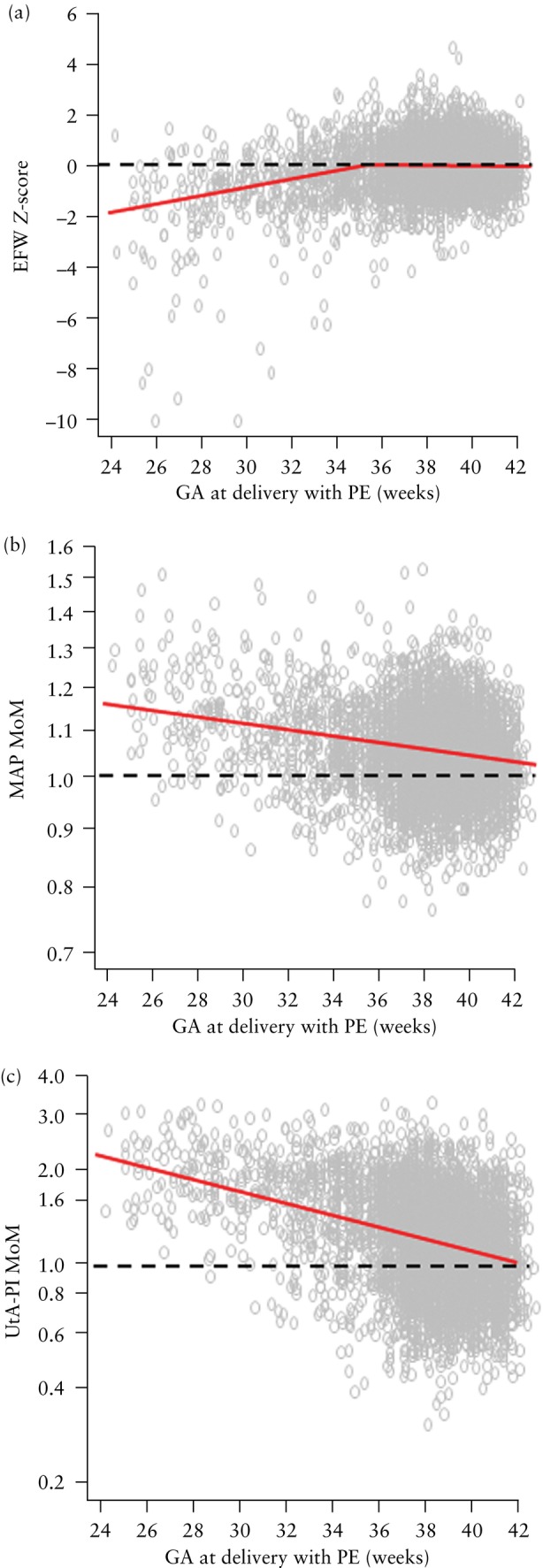
Objective: To stratify pregnancy care based on the estimated risk of pre-eclampsia (PE) from screening at 19-24 weeks' gestation by combinations of maternal risk factors, estimated fetal weight (EFW), mean arterial pressure (MAP) and uterine artery pulsatility index (UtA-PI).

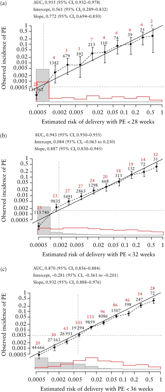
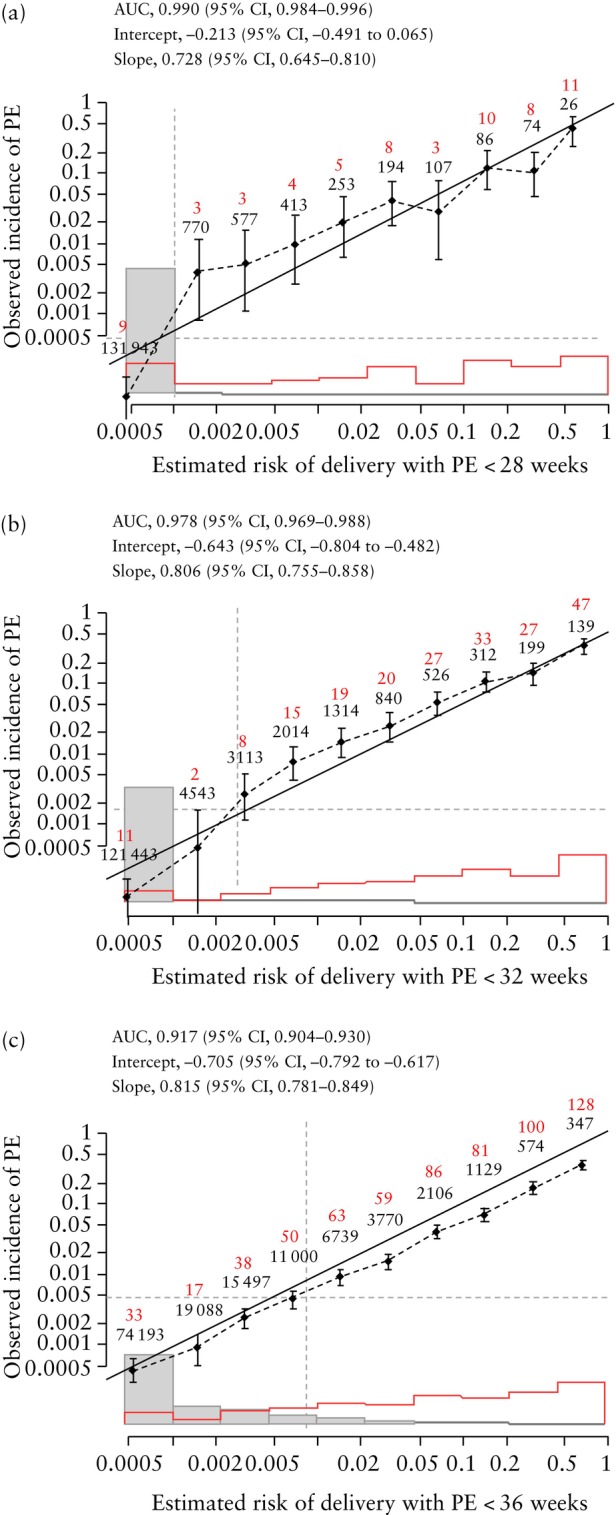
Methods: The data for this study were derived from a prospective non-interventional study in 134 443 women with a singleton pregnancy attending for a routine ultrasound scan at 19 + 0 to 23 + 6 weeks' gestation in two UK maternity hospitals. The visit included recording of maternal demographic characteristics and medical history, sonographic EFW and measurement of MAP and UtA-PI. The competing-risks model was used to estimate the individual patient-specific risk of delivery with PE at < 28, < 32 and < 36 weeks' gestation. Receiver-operating-characteristics curves were constructed for screen-positive rates (SPRs) at different detection rates of delivery with PE at < 28, < 32 and < 36 weeks' gestation for the combinations of maternal risk factors, EFW and MAP, and of maternal risk factors, EFW, MAP and UtA-PI. Different risk cut-offs were used with the intention of detecting about 80%, 85% and 90% of cases of delivery with PE at < 28, < 32 and < 36 weeks' gestation. Calibration for risk of delivery with PE at < 28, < 32 and < 36 weeks' gestation was assessed by plotting the observed incidence of PE against the predicted incidence of PE.

Results: The study population contained 4335 (3.2%) women that subsequently developed PE, including 64 (0.05%) that delivered with PE at < 28 weeks' gestation, 209 (0.2%) that delivered with PE at < 32 weeks and 655 (0.5%) that delivered with PE at < 36 weeks. If the objective of screening was to identify about 90% of cases of delivery with PE at < 28, < 32 and < 36 weeks and the method of screening was a combination of maternal risk factors, EFW and MAP, the respective SPRs would be 11.0%, 18.3% and 38.8%. If the method of screening also included UtA-PI, the respective SPRs would be 2.6%, 3.8% and 23.6%. If the objective of screening was to identify about 80% of cases of delivery with PE at < 28, < 32 and < 36 weeks and the method of screening was a combination of maternal risk factors, EFW and MAP, the respective SPRs would be 5.9%, 9.7% and 21.9%. If the method of screening also included UtA-PI, the respective SPRs would be 1.0%, 2.1% and 11.7%. The calibration plots demonstrated good agreement between the estimated risk and observed incidence of PE.

Conclusions: All women should be offered assessment of risk for PE at 11-13 weeks, to help identify those requiring aspirin prophylaxis to reduce the rate of preterm PE, and at 35-37 weeks, to determine the optimal timing of birth to reduce the rate of term PE. Assessment of risk for PE at mid-gestation can be used to identify the subgroups that require additional monitoring at 24-35, 28-35 and 32-35 weeks' gestation. The best performance of screening, reflected in the SPR necessary to achieve a high detection rate, is achieved by a combination of maternal risk factors, MAP and UtA-PI. © 2025 The Author(s). Ultrasound in Obstetrics & Gynecology published by John Wiley & Sons Ltd on behalf of International Society of Ultrasound in Obstetrics and Gynecology.

求助全文
通过发布文献求助,成功后即可免费获取论文全文。 去求助
来源期刊
CiteScore
12.30
自引率
14.10%
发文量
891
审稿时长
1 months
期刊介绍: Ultrasound in Obstetrics & Gynecology (UOG) is the official journal of the International Society of Ultrasound in Obstetrics and Gynecology (ISUOG) and is considered the foremost international peer-reviewed journal in the field. It publishes cutting-edge research that is highly relevant to clinical practice, which includes guidelines, expert commentaries, consensus statements, original articles, and systematic reviews. UOG is widely recognized and included in prominent abstract and indexing databases such as Index Medicus and Current Contents.
×
引用
GB/T 7714-2015
复制
MLA
复制
APA
复制
导出至
BibTeX EndNote RefMan NoteFirst NoteExpress
×
提示
您的信息不完整,为了账户安全,请先补充。
现在去补充
×
提示
您因"违规操作"
具体请查看互助需知
我知道了
×
提示
确定
请完成安全验证×
copy
已复制链接
快去分享给好友吧!
我知道了
右上角分享
点击右上角分享
0
联系我们:info@booksci.cn Book学术提供免费学术资源搜索服务,方便国内外学者检索中英文文献。致力于提供最便捷和优质的服务体验。 Copyright © 2023 布克学术 All rights reserved.
京ICP备2023020795号-1
ghs 京公网安备 11010802042870号
Book学术文献互助
Book学术文献互助群
群 号:604180095
Book学术官方微信