{"title":"继发于糖尿病足溃疡的软组织及骨内肺肿:一例严重的肺气肿性骨髓炎。","authors":"Emmanuel Olayinka Sobamowo, Mirza Shaheer Baig, Sumantra Kumar, Nikhil Rasik Patel","doi":"10.1093/bjrcr/uaaf016","DOIUrl":null,"url":null,"abstract":"<p><p>Emphysematous osteomyelitis (EO) is an uncommon but severe form of osteomyelitis that is characterized by gas formation within the bone. This case report highlights a case of particularly severe EO in an amputated foot, with key imaging findings across modalities emphasizing the diagnostic challenges and the importance of early detection. A 68-year-old male with a history of poorly controlled diabetes and a previous left third to fifth toe amputation for a non-healing ulcer presented to the emergency department with an infective picture and poorly controlled blood glucose levels. After clinical assessment, a focus of infection was found in the left foot and was subsequently assessed with plain radiography, MRI, and CT. The case highlighted the utility of each modality in such a complex presentation, including trabecular bony changes on the plain radiograph, soft tissue changes on MRI and confirmation of intraosseous pneumatosis on CT. This case highlights key imaging features of EO and underscores the need to use CT and MRI to guide timely surgical and medical management. This report adds to the limited literature on EO and presents a useful acronym of \"LEAP\" to describe key features when suspecting EO - lack of cortical destruction, extra-osseous soft tissue gas, associated comorbidities (diabetes, malignancy, etc), and pumice stone sign.</p>","PeriodicalId":45216,"journal":{"name":"BJR Case Reports","volume":"11 3","pages":"uaaf016"},"PeriodicalIF":0.5000,"publicationDate":"2025-03-18","publicationTypes":"Journal Article","fieldsOfStudy":null,"isOpenAccess":false,"openAccessPdf":"https://www.ncbi.nlm.nih.gov/pmc/articles/PMC12048179/pdf/","citationCount":"0","resultStr":"{\"title\":\"Soft tissue and intraosseous pneumatosis secondary to diabetic foot ulcer: a severe case of emphysematous osteomyelitis.\",\"authors\":\"Emmanuel Olayinka Sobamowo, Mirza Shaheer Baig, Sumantra Kumar, Nikhil Rasik Patel\",\"doi\":\"10.1093/bjrcr/uaaf016\",\"DOIUrl\":null,\"url\":null,\"abstract\":\"<p><p>Emphysematous osteomyelitis (EO) is an uncommon but severe form of osteomyelitis that is characterized by gas formation within the bone. This case report highlights a case of particularly severe EO in an amputated foot, with key imaging findings across modalities emphasizing the diagnostic challenges and the importance of early detection. A 68-year-old male with a history of poorly controlled diabetes and a previous left third to fifth toe amputation for a non-healing ulcer presented to the emergency department with an infective picture and poorly controlled blood glucose levels. After clinical assessment, a focus of infection was found in the left foot and was subsequently assessed with plain radiography, MRI, and CT. The case highlighted the utility of each modality in such a complex presentation, including trabecular bony changes on the plain radiograph, soft tissue changes on MRI and confirmation of intraosseous pneumatosis on CT. This case highlights key imaging features of EO and underscores the need to use CT and MRI to guide timely surgical and medical management. This report adds to the limited literature on EO and presents a useful acronym of \\\"LEAP\\\" to describe key features when suspecting EO - lack of cortical destruction, extra-osseous soft tissue gas, associated comorbidities (diabetes, malignancy, etc), and pumice stone sign.</p>\",\"PeriodicalId\":45216,\"journal\":{\"name\":\"BJR Case Reports\",\"volume\":\"11 3\",\"pages\":\"uaaf016\"},\"PeriodicalIF\":0.5000,\"publicationDate\":\"2025-03-18\",\"publicationTypes\":\"Journal Article\",\"fieldsOfStudy\":null,\"isOpenAccess\":false,\"openAccessPdf\":\"https://www.ncbi.nlm.nih.gov/pmc/articles/PMC12048179/pdf/\",\"citationCount\":\"0\",\"resultStr\":null,\"platform\":\"Semanticscholar\",\"paperid\":null,\"PeriodicalName\":\"BJR Case Reports\",\"FirstCategoryId\":\"1085\",\"ListUrlMain\":\"https://doi.org/10.1093/bjrcr/uaaf016\",\"RegionNum\":0,\"RegionCategory\":null,\"ArticlePicture\":[],\"TitleCN\":null,\"AbstractTextCN\":null,\"PMCID\":null,\"EPubDate\":\"2025/5/1 0:00:00\",\"PubModel\":\"eCollection\",\"JCR\":\"Q4\",\"JCRName\":\"RADIOLOGY, NUCLEAR MEDICINE & MEDICAL IMAGING\",\"Score\":null,\"Total\":0}","platform":"Semanticscholar","paperid":null,"PeriodicalName":"BJR Case Reports","FirstCategoryId":"1085","ListUrlMain":"https://doi.org/10.1093/bjrcr/uaaf016","RegionNum":0,"RegionCategory":null,"ArticlePicture":[],"TitleCN":null,"AbstractTextCN":null,"PMCID":null,"EPubDate":"2025/5/1 0:00:00","PubModel":"eCollection","JCR":"Q4","JCRName":"RADIOLOGY, NUCLEAR MEDICINE & MEDICAL IMAGING","Score":null,"Total":0}
Soft tissue and intraosseous pneumatosis secondary to diabetic foot ulcer: a severe case of emphysematous osteomyelitis.
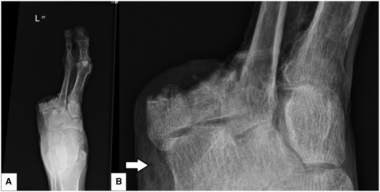
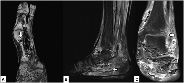
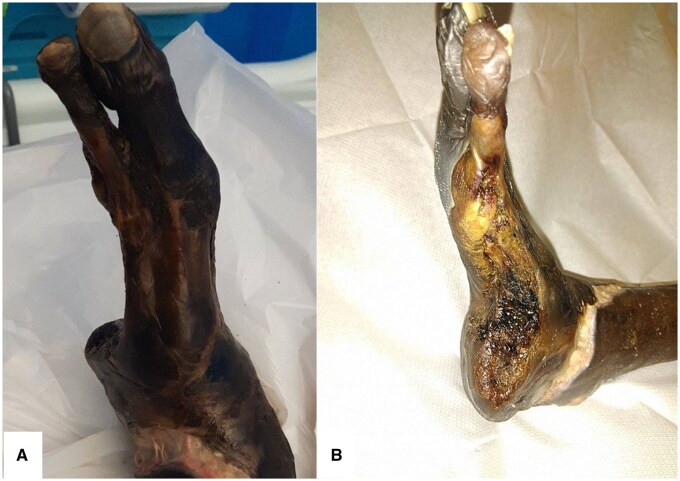
Emphysematous osteomyelitis (EO) is an uncommon but severe form of osteomyelitis that is characterized by gas formation within the bone. This case report highlights a case of particularly severe EO in an amputated foot, with key imaging findings across modalities emphasizing the diagnostic challenges and the importance of early detection. A 68-year-old male with a history of poorly controlled diabetes and a previous left third to fifth toe amputation for a non-healing ulcer presented to the emergency department with an infective picture and poorly controlled blood glucose levels. After clinical assessment, a focus of infection was found in the left foot and was subsequently assessed with plain radiography, MRI, and CT. The case highlighted the utility of each modality in such a complex presentation, including trabecular bony changes on the plain radiograph, soft tissue changes on MRI and confirmation of intraosseous pneumatosis on CT. This case highlights key imaging features of EO and underscores the need to use CT and MRI to guide timely surgical and medical management. This report adds to the limited literature on EO and presents a useful acronym of "LEAP" to describe key features when suspecting EO - lack of cortical destruction, extra-osseous soft tissue gas, associated comorbidities (diabetes, malignancy, etc), and pumice stone sign.

 求助内容:
求助内容: 应助结果提醒方式:


