Abdulaziz Saber, Mohammed Khashab, Abdulhadi Turkistani, Moyassar Karami, Saleh Almaymoni, Mohamed Elkhalifa, Abdulhadi Algahtani, Abdullah Alhazmi
{"title":"三维导航引导下手术切除罕见的骶骨间充质软骨肉瘤1例。","authors":"Abdulaziz Saber, Mohammed Khashab, Abdulhadi Turkistani, Moyassar Karami, Saleh Almaymoni, Mohamed Elkhalifa, Abdulhadi Algahtani, Abdullah Alhazmi","doi":"10.21037/jss-24-104","DOIUrl":null,"url":null,"abstract":"<p><strong>Background: </strong>Chondrosarcomas are a group of heterogeneous malignant cartilaginous neoplasms that arise from preexisting benign precursors. They can be divided into conventional (primary) chondrosarcomas, which account for 90% of cases, and nonconventional chondrosarcomas, which account for the remaining 10%. Mesenchymal chondrosarcoma (MCS) is a rare high-grade soft tissue tumor variant of nonconventional chondrosarcoma that is histologically characterized by a biphasic pattern of atypical cartilage with small round cells.</p><p><strong>Case description: </strong>A 23-year-old female known case of ovarian cyst presented with a two-year history of low back pain and constitutional symptoms. Pelvic magnetic resonance imaging (MRI) with contrast showed a well-defined lesion with intermediate to high signals located at the right wing of the upper sacrum, at the level of S1-S2. The patient underwent a combined ultrasound and computed tomography-guided biopsy under local anesthesia and the immunochemical profile was positive for CD99 and S100 biomarkers. The patient underwent a two-stage procedure for a wide marginal tumor resection. Stage 1 was performed with an anterior approach; identification of the tumor margins was done followed by designing the cuts of the sacrum to achieve wide margins around the tumor. Stage 2 was performed with a posterior approach exposing L3 vertebrae down to the sacrum. Utilizing O-Arm Navigation for posterior margin allocation in addition to instrumentation. After 12 months post-operation, follow up revealed no evidence of recurrence.</p><p><strong>Conclusions: </strong>Limitation in accessibility to the axial skeleton and the neurovascular component, poses a challenge to treatment. Therefore, using neuro-navigation system and optimal adjuvant therapy should be studied further to improve the prognosis.</p>","PeriodicalId":17131,"journal":{"name":"Journal of spine surgery","volume":"11 1","pages":"197-205"},"PeriodicalIF":0.0000,"publicationDate":"2025-03-24","publicationTypes":"Journal Article","fieldsOfStudy":null,"isOpenAccess":false,"openAccessPdf":"https://www.ncbi.nlm.nih.gov/pmc/articles/PMC11998042/pdf/","citationCount":"0","resultStr":"{\"title\":\"A 3D navigation-guided surgical resection of a rare case of sacral spine mesenchymal chondrosarcoma: a case report.\",\"authors\":\"Abdulaziz Saber, Mohammed Khashab, Abdulhadi Turkistani, Moyassar Karami, Saleh Almaymoni, Mohamed Elkhalifa, Abdulhadi Algahtani, Abdullah Alhazmi\",\"doi\":\"10.21037/jss-24-104\",\"DOIUrl\":null,\"url\":null,\"abstract\":\"<p><strong>Background: </strong>Chondrosarcomas are a group of heterogeneous malignant cartilaginous neoplasms that arise from preexisting benign precursors. They can be divided into conventional (primary) chondrosarcomas, which account for 90% of cases, and nonconventional chondrosarcomas, which account for the remaining 10%. Mesenchymal chondrosarcoma (MCS) is a rare high-grade soft tissue tumor variant of nonconventional chondrosarcoma that is histologically characterized by a biphasic pattern of atypical cartilage with small round cells.</p><p><strong>Case description: </strong>A 23-year-old female known case of ovarian cyst presented with a two-year history of low back pain and constitutional symptoms. Pelvic magnetic resonance imaging (MRI) with contrast showed a well-defined lesion with intermediate to high signals located at the right wing of the upper sacrum, at the level of S1-S2. The patient underwent a combined ultrasound and computed tomography-guided biopsy under local anesthesia and the immunochemical profile was positive for CD99 and S100 biomarkers. The patient underwent a two-stage procedure for a wide marginal tumor resection. Stage 1 was performed with an anterior approach; identification of the tumor margins was done followed by designing the cuts of the sacrum to achieve wide margins around the tumor. Stage 2 was performed with a posterior approach exposing L3 vertebrae down to the sacrum. Utilizing O-Arm Navigation for posterior margin allocation in addition to instrumentation. After 12 months post-operation, follow up revealed no evidence of recurrence.</p><p><strong>Conclusions: </strong>Limitation in accessibility to the axial skeleton and the neurovascular component, poses a challenge to treatment. Therefore, using neuro-navigation system and optimal adjuvant therapy should be studied further to improve the prognosis.</p>\",\"PeriodicalId\":17131,\"journal\":{\"name\":\"Journal of spine surgery\",\"volume\":\"11 1\",\"pages\":\"197-205\"},\"PeriodicalIF\":0.0000,\"publicationDate\":\"2025-03-24\",\"publicationTypes\":\"Journal Article\",\"fieldsOfStudy\":null,\"isOpenAccess\":false,\"openAccessPdf\":\"https://www.ncbi.nlm.nih.gov/pmc/articles/PMC11998042/pdf/\",\"citationCount\":\"0\",\"resultStr\":null,\"platform\":\"Semanticscholar\",\"paperid\":null,\"PeriodicalName\":\"Journal of spine surgery\",\"FirstCategoryId\":\"1085\",\"ListUrlMain\":\"https://doi.org/10.21037/jss-24-104\",\"RegionNum\":0,\"RegionCategory\":null,\"ArticlePicture\":[],\"TitleCN\":null,\"AbstractTextCN\":null,\"PMCID\":null,\"EPubDate\":\"2025/3/11 0:00:00\",\"PubModel\":\"Epub\",\"JCR\":\"Q1\",\"JCRName\":\"Medicine\",\"Score\":null,\"Total\":0}","platform":"Semanticscholar","paperid":null,"PeriodicalName":"Journal of spine surgery","FirstCategoryId":"1085","ListUrlMain":"https://doi.org/10.21037/jss-24-104","RegionNum":0,"RegionCategory":null,"ArticlePicture":[],"TitleCN":null,"AbstractTextCN":null,"PMCID":null,"EPubDate":"2025/3/11 0:00:00","PubModel":"Epub","JCR":"Q1","JCRName":"Medicine","Score":null,"Total":0}
A 3D navigation-guided surgical resection of a rare case of sacral spine mesenchymal chondrosarcoma: a case report.
Background: Chondrosarcomas are a group of heterogeneous malignant cartilaginous neoplasms that arise from preexisting benign precursors. They can be divided into conventional (primary) chondrosarcomas, which account for 90% of cases, and nonconventional chondrosarcomas, which account for the remaining 10%. Mesenchymal chondrosarcoma (MCS) is a rare high-grade soft tissue tumor variant of nonconventional chondrosarcoma that is histologically characterized by a biphasic pattern of atypical cartilage with small round cells.
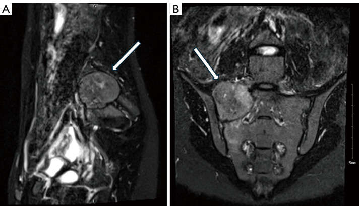
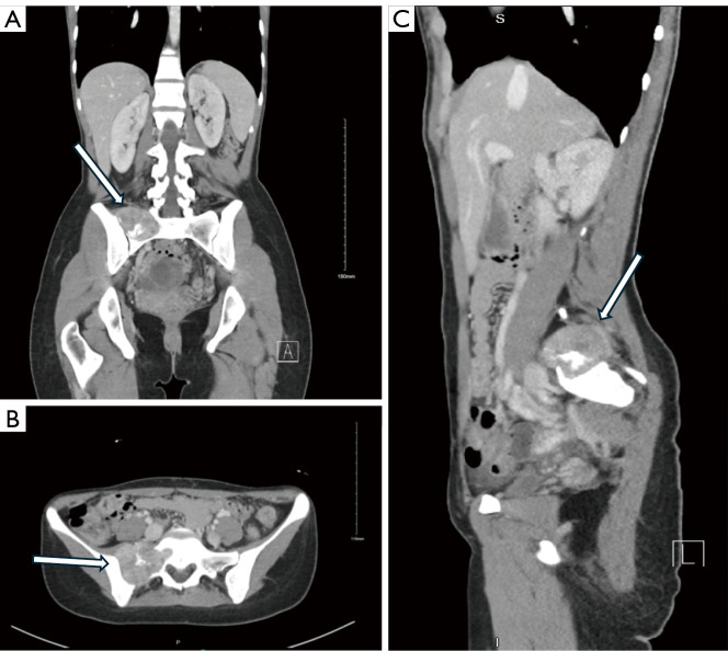
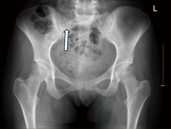
Case description: A 23-year-old female known case of ovarian cyst presented with a two-year history of low back pain and constitutional symptoms. Pelvic magnetic resonance imaging (MRI) with contrast showed a well-defined lesion with intermediate to high signals located at the right wing of the upper sacrum, at the level of S1-S2. The patient underwent a combined ultrasound and computed tomography-guided biopsy under local anesthesia and the immunochemical profile was positive for CD99 and S100 biomarkers. The patient underwent a two-stage procedure for a wide marginal tumor resection. Stage 1 was performed with an anterior approach; identification of the tumor margins was done followed by designing the cuts of the sacrum to achieve wide margins around the tumor. Stage 2 was performed with a posterior approach exposing L3 vertebrae down to the sacrum. Utilizing O-Arm Navigation for posterior margin allocation in addition to instrumentation. After 12 months post-operation, follow up revealed no evidence of recurrence.
Conclusions: Limitation in accessibility to the axial skeleton and the neurovascular component, poses a challenge to treatment. Therefore, using neuro-navigation system and optimal adjuvant therapy should be studied further to improve the prognosis.

 求助内容:
求助内容: 应助结果提醒方式:


