Elena Zoppolato, Hasse Mol, Carlos Estrella-García, Nicole Vizcaino-Rodríguez, Diana Sanchez, Nicole Procel, Isabel Baroja, Leticia Sansores-Garcia, Iván M Moya
{"title":"优化免疫荧光肝脏结构分析:提高三维分辨率和最大限度地减少组织自身荧光。","authors":"Elena Zoppolato, Hasse Mol, Carlos Estrella-García, Nicole Vizcaino-Rodríguez, Diana Sanchez, Nicole Procel, Isabel Baroja, Leticia Sansores-Garcia, Iván M Moya","doi":"10.1093/biomethods/bpaf023","DOIUrl":null,"url":null,"abstract":"<p><p>The study of liver biology and pathology through marker expression analysis and tissue structure visualization is constrained by the high autofluorescence caused by the presence of lipofuscins, vitamin A, and lipid droplets, which traditional staining methods do not effectively quench. This leads to low signal-to-noise ratios, obscured expression levels, and reduced structural resolution. We mitigated liver tissue autofluorescence using Sudan Black B staining, which effectively quenches background signals from lipid and lipofuscin accumulation. Additionally, these protocols typically use thin paraffin sections (5-7 µm), which limit the analysis of larger and more complex liver structures. Liver tissue is highly organized in three dimensions, with large hepatocytes (20-30 µm in diameter) arranged around sinusoids and bile canaliculi, which form intricate branching networks. Thin sections cannot capture this 3D organization, providing only a \"snapshot\" of the tissue at one plane. Here, we present an optimized immunofluorescence protocol using 100-200 µm vibratome-cut liver sections to enable a more comprehensive 3D-like analysis of liver architecture. Finally, our protocol includes antigen retrieval steps tailored to each antibody, maximizing epitope accessibility and signal clarity. Together, these improvements provide a robust method for detailed liver studies with enhanced specificity and structural resolution in immunofluorescent staining. This protocol is particularly suited for researchers focused on liver regeneration, cancer, chronic disease pathology, and structural analysis. However, other researchers interested in exploring complex tissue structures in other autofluorescent tissues, such as the kidney, brain, pancreas, spleen, and adipose tissue, will also find this method beneficial.</p>","PeriodicalId":36528,"journal":{"name":"Biology Methods and Protocols","volume":"10 1","pages":"bpaf023"},"PeriodicalIF":1.3000,"publicationDate":"2025-03-26","publicationTypes":"Journal Article","fieldsOfStudy":null,"isOpenAccess":false,"openAccessPdf":"https://www.ncbi.nlm.nih.gov/pmc/articles/PMC11999924/pdf/","citationCount":"0","resultStr":"{\"title\":\"Optimized immunofluorescence for liver structure analysis: Enhancing 3D resolution and minimizing tissue autofluorescence.\",\"authors\":\"Elena Zoppolato, Hasse Mol, Carlos Estrella-García, Nicole Vizcaino-Rodríguez, Diana Sanchez, Nicole Procel, Isabel Baroja, Leticia Sansores-Garcia, Iván M Moya\",\"doi\":\"10.1093/biomethods/bpaf023\",\"DOIUrl\":null,\"url\":null,\"abstract\":\"<p><p>The study of liver biology and pathology through marker expression analysis and tissue structure visualization is constrained by the high autofluorescence caused by the presence of lipofuscins, vitamin A, and lipid droplets, which traditional staining methods do not effectively quench. This leads to low signal-to-noise ratios, obscured expression levels, and reduced structural resolution. We mitigated liver tissue autofluorescence using Sudan Black B staining, which effectively quenches background signals from lipid and lipofuscin accumulation. Additionally, these protocols typically use thin paraffin sections (5-7 µm), which limit the analysis of larger and more complex liver structures. Liver tissue is highly organized in three dimensions, with large hepatocytes (20-30 µm in diameter) arranged around sinusoids and bile canaliculi, which form intricate branching networks. Thin sections cannot capture this 3D organization, providing only a \\\"snapshot\\\" of the tissue at one plane. Here, we present an optimized immunofluorescence protocol using 100-200 µm vibratome-cut liver sections to enable a more comprehensive 3D-like analysis of liver architecture. Finally, our protocol includes antigen retrieval steps tailored to each antibody, maximizing epitope accessibility and signal clarity. Together, these improvements provide a robust method for detailed liver studies with enhanced specificity and structural resolution in immunofluorescent staining. This protocol is particularly suited for researchers focused on liver regeneration, cancer, chronic disease pathology, and structural analysis. However, other researchers interested in exploring complex tissue structures in other autofluorescent tissues, such as the kidney, brain, pancreas, spleen, and adipose tissue, will also find this method beneficial.</p>\",\"PeriodicalId\":36528,\"journal\":{\"name\":\"Biology Methods and Protocols\",\"volume\":\"10 1\",\"pages\":\"bpaf023\"},\"PeriodicalIF\":1.3000,\"publicationDate\":\"2025-03-26\",\"publicationTypes\":\"Journal Article\",\"fieldsOfStudy\":null,\"isOpenAccess\":false,\"openAccessPdf\":\"https://www.ncbi.nlm.nih.gov/pmc/articles/PMC11999924/pdf/\",\"citationCount\":\"0\",\"resultStr\":null,\"platform\":\"Semanticscholar\",\"paperid\":null,\"PeriodicalName\":\"Biology Methods and Protocols\",\"FirstCategoryId\":\"1085\",\"ListUrlMain\":\"https://doi.org/10.1093/biomethods/bpaf023\",\"RegionNum\":0,\"RegionCategory\":null,\"ArticlePicture\":[],\"TitleCN\":null,\"AbstractTextCN\":null,\"PMCID\":null,\"EPubDate\":\"2025/1/1 0:00:00\",\"PubModel\":\"eCollection\",\"JCR\":\"Q3\",\"JCRName\":\"BIOCHEMICAL RESEARCH METHODS\",\"Score\":null,\"Total\":0}","platform":"Semanticscholar","paperid":null,"PeriodicalName":"Biology Methods and Protocols","FirstCategoryId":"1085","ListUrlMain":"https://doi.org/10.1093/biomethods/bpaf023","RegionNum":0,"RegionCategory":null,"ArticlePicture":[],"TitleCN":null,"AbstractTextCN":null,"PMCID":null,"EPubDate":"2025/1/1 0:00:00","PubModel":"eCollection","JCR":"Q3","JCRName":"BIOCHEMICAL RESEARCH METHODS","Score":null,"Total":0}
Optimized immunofluorescence for liver structure analysis: Enhancing 3D resolution and minimizing tissue autofluorescence.
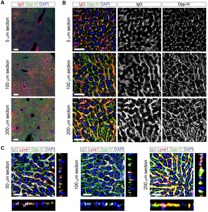
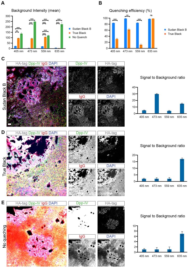
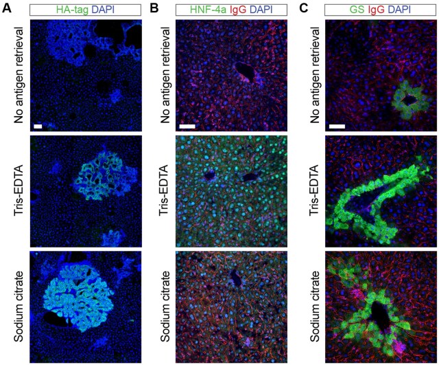
The study of liver biology and pathology through marker expression analysis and tissue structure visualization is constrained by the high autofluorescence caused by the presence of lipofuscins, vitamin A, and lipid droplets, which traditional staining methods do not effectively quench. This leads to low signal-to-noise ratios, obscured expression levels, and reduced structural resolution. We mitigated liver tissue autofluorescence using Sudan Black B staining, which effectively quenches background signals from lipid and lipofuscin accumulation. Additionally, these protocols typically use thin paraffin sections (5-7 µm), which limit the analysis of larger and more complex liver structures. Liver tissue is highly organized in three dimensions, with large hepatocytes (20-30 µm in diameter) arranged around sinusoids and bile canaliculi, which form intricate branching networks. Thin sections cannot capture this 3D organization, providing only a "snapshot" of the tissue at one plane. Here, we present an optimized immunofluorescence protocol using 100-200 µm vibratome-cut liver sections to enable a more comprehensive 3D-like analysis of liver architecture. Finally, our protocol includes antigen retrieval steps tailored to each antibody, maximizing epitope accessibility and signal clarity. Together, these improvements provide a robust method for detailed liver studies with enhanced specificity and structural resolution in immunofluorescent staining. This protocol is particularly suited for researchers focused on liver regeneration, cancer, chronic disease pathology, and structural analysis. However, other researchers interested in exploring complex tissue structures in other autofluorescent tissues, such as the kidney, brain, pancreas, spleen, and adipose tissue, will also find this method beneficial.

 求助内容:
求助内容: 应助结果提醒方式:


