{"title":"股骨皮下囊肿后发生的假体周围关节感染:全膝关节置换术后罕见的并发症。","authors":"Naohiro Oka, Shigeshi Mori, Yu Shinyashiki, Nobuhisa Shokaku, Kenji Yamazaki, Koji Goto, Daisuke Togawa","doi":"10.1155/cro/7710384","DOIUrl":null,"url":null,"abstract":"<p><p>Herein, we present a rare case of periprosthetic joint infection (PJI) which was triggered by an infection with a latent subcutaneous cyst on the thigh and occurred in a strange course following total knee arthroplasty (TKA). An 87-year-old female underwent right TKA followed by left TKA 5 months later. Six weeks after left TKA, a painful subcutaneous induration appeared in the left medial thigh. Magnetic resonance imaging revealed a 30∗50-mm multifocal mass. The cystic fluid was brown and cloudy, indicating an infected cyst. Oral antimicrobial therapy was initiated for 7 days. Nine weeks after the left TKA, a left calcaneal fracture occurred. Subsequently, edema of the lower extremities and pain in the left knee gradually developed. Arthrocentesis was performed twice: joint fluid Gram staining and culture examination were negative. However, at 12.5 weeks, an alpha-defensin test of the synovial fluid was positive. Therefore, PJI was diagnosed. DAIR was performed, followed by multiantibiotic therapy. The infection subsided gradually. Edema of the lower limbs was treated with oral diuretics, lymphatic massage, and compression stockings. Consequently, the lower limb edema also improved. In this case, infection of a latent subcutaneous cyst in the thigh occurred and spread around the prosthesis due to leg edema, which was associated with loss of lower limb motion due to a calcaneal fracture. The presence of a potential thigh subcutaneous cyst is a risk factor for PJI. Moreover, lower extremity edema occurs by decreasing lower extremity motion, such as after a calcaneal fracture, and it increases the risk of extending extra-articular infection to the PJI. Potential thigh subcutaneous cysts and lower extremity edema are risk factors for the development of PJI. Orthopedic surgeons need to be aware of these facts during follow-up after TKA.</p>","PeriodicalId":30287,"journal":{"name":"Case Reports in Orthopedics","volume":"2025 ","pages":"7710384"},"PeriodicalIF":0.6000,"publicationDate":"2025-04-24","publicationTypes":"Journal Article","fieldsOfStudy":null,"isOpenAccess":false,"openAccessPdf":"https://www.ncbi.nlm.nih.gov/pmc/articles/PMC12045695/pdf/","citationCount":"0","resultStr":"{\"title\":\"Periprosthetic Joint Infection Occurring Following a Femoral Subcutaneous Cyst: A Rare Complication Post-Total Knee Arthroplasty.\",\"authors\":\"Naohiro Oka, Shigeshi Mori, Yu Shinyashiki, Nobuhisa Shokaku, Kenji Yamazaki, Koji Goto, Daisuke Togawa\",\"doi\":\"10.1155/cro/7710384\",\"DOIUrl\":null,\"url\":null,\"abstract\":\"<p><p>Herein, we present a rare case of periprosthetic joint infection (PJI) which was triggered by an infection with a latent subcutaneous cyst on the thigh and occurred in a strange course following total knee arthroplasty (TKA). An 87-year-old female underwent right TKA followed by left TKA 5 months later. Six weeks after left TKA, a painful subcutaneous induration appeared in the left medial thigh. Magnetic resonance imaging revealed a 30∗50-mm multifocal mass. The cystic fluid was brown and cloudy, indicating an infected cyst. Oral antimicrobial therapy was initiated for 7 days. Nine weeks after the left TKA, a left calcaneal fracture occurred. Subsequently, edema of the lower extremities and pain in the left knee gradually developed. Arthrocentesis was performed twice: joint fluid Gram staining and culture examination were negative. However, at 12.5 weeks, an alpha-defensin test of the synovial fluid was positive. Therefore, PJI was diagnosed. DAIR was performed, followed by multiantibiotic therapy. The infection subsided gradually. Edema of the lower limbs was treated with oral diuretics, lymphatic massage, and compression stockings. Consequently, the lower limb edema also improved. In this case, infection of a latent subcutaneous cyst in the thigh occurred and spread around the prosthesis due to leg edema, which was associated with loss of lower limb motion due to a calcaneal fracture. The presence of a potential thigh subcutaneous cyst is a risk factor for PJI. Moreover, lower extremity edema occurs by decreasing lower extremity motion, such as after a calcaneal fracture, and it increases the risk of extending extra-articular infection to the PJI. Potential thigh subcutaneous cysts and lower extremity edema are risk factors for the development of PJI. Orthopedic surgeons need to be aware of these facts during follow-up after TKA.</p>\",\"PeriodicalId\":30287,\"journal\":{\"name\":\"Case Reports in Orthopedics\",\"volume\":\"2025 \",\"pages\":\"7710384\"},\"PeriodicalIF\":0.6000,\"publicationDate\":\"2025-04-24\",\"publicationTypes\":\"Journal Article\",\"fieldsOfStudy\":null,\"isOpenAccess\":false,\"openAccessPdf\":\"https://www.ncbi.nlm.nih.gov/pmc/articles/PMC12045695/pdf/\",\"citationCount\":\"0\",\"resultStr\":null,\"platform\":\"Semanticscholar\",\"paperid\":null,\"PeriodicalName\":\"Case Reports in Orthopedics\",\"FirstCategoryId\":\"1085\",\"ListUrlMain\":\"https://doi.org/10.1155/cro/7710384\",\"RegionNum\":0,\"RegionCategory\":null,\"ArticlePicture\":[],\"TitleCN\":null,\"AbstractTextCN\":null,\"PMCID\":null,\"EPubDate\":\"2025/1/1 0:00:00\",\"PubModel\":\"eCollection\",\"JCR\":\"Q4\",\"JCRName\":\"ORTHOPEDICS\",\"Score\":null,\"Total\":0}","platform":"Semanticscholar","paperid":null,"PeriodicalName":"Case Reports in Orthopedics","FirstCategoryId":"1085","ListUrlMain":"https://doi.org/10.1155/cro/7710384","RegionNum":0,"RegionCategory":null,"ArticlePicture":[],"TitleCN":null,"AbstractTextCN":null,"PMCID":null,"EPubDate":"2025/1/1 0:00:00","PubModel":"eCollection","JCR":"Q4","JCRName":"ORTHOPEDICS","Score":null,"Total":0}
Periprosthetic Joint Infection Occurring Following a Femoral Subcutaneous Cyst: A Rare Complication Post-Total Knee Arthroplasty.
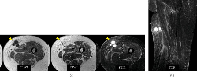
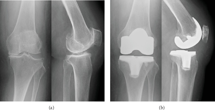
Herein, we present a rare case of periprosthetic joint infection (PJI) which was triggered by an infection with a latent subcutaneous cyst on the thigh and occurred in a strange course following total knee arthroplasty (TKA). An 87-year-old female underwent right TKA followed by left TKA 5 months later. Six weeks after left TKA, a painful subcutaneous induration appeared in the left medial thigh. Magnetic resonance imaging revealed a 30∗50-mm multifocal mass. The cystic fluid was brown and cloudy, indicating an infected cyst. Oral antimicrobial therapy was initiated for 7 days. Nine weeks after the left TKA, a left calcaneal fracture occurred. Subsequently, edema of the lower extremities and pain in the left knee gradually developed. Arthrocentesis was performed twice: joint fluid Gram staining and culture examination were negative. However, at 12.5 weeks, an alpha-defensin test of the synovial fluid was positive. Therefore, PJI was diagnosed. DAIR was performed, followed by multiantibiotic therapy. The infection subsided gradually. Edema of the lower limbs was treated with oral diuretics, lymphatic massage, and compression stockings. Consequently, the lower limb edema also improved. In this case, infection of a latent subcutaneous cyst in the thigh occurred and spread around the prosthesis due to leg edema, which was associated with loss of lower limb motion due to a calcaneal fracture. The presence of a potential thigh subcutaneous cyst is a risk factor for PJI. Moreover, lower extremity edema occurs by decreasing lower extremity motion, such as after a calcaneal fracture, and it increases the risk of extending extra-articular infection to the PJI. Potential thigh subcutaneous cysts and lower extremity edema are risk factors for the development of PJI. Orthopedic surgeons need to be aware of these facts during follow-up after TKA.


 求助内容:
求助内容: 应助结果提醒方式:


