Hosamadin S Assadi, Xiaodan Zhao, Gareth Matthews, Rui Li, Jordi Broncano Cabrero, Bahman Kasmai, Samer Alabed, Javier Royuela Del Val, Hilmar Spohr, Yashoda Gurung-Koney, Nay Aung, Sunil Nair, Andrew J Swift, Vassilios S Vassiliou, Liang Zhong, Abdallah Al-Mohammad, Rob J van der Geest, Peter P Swoboda, Sven Plein, Pankaj Garg
{"title":"衰老的心血管磁共振成像标志物:一项多中心、横断面队列研究。","authors":"Hosamadin S Assadi, Xiaodan Zhao, Gareth Matthews, Rui Li, Jordi Broncano Cabrero, Bahman Kasmai, Samer Alabed, Javier Royuela Del Val, Hilmar Spohr, Yashoda Gurung-Koney, Nay Aung, Sunil Nair, Andrew J Swift, Vassilios S Vassiliou, Liang Zhong, Abdallah Al-Mohammad, Rob J van der Geest, Peter P Swoboda, Sven Plein, Pankaj Garg","doi":"10.1093/ehjopen/oeaf032","DOIUrl":null,"url":null,"abstract":"<p><strong>Aims: </strong>Cardiac ageing involves a series of anatomical and physiological changes contributing to a decline in overall performance. Cardiac magnetic resonance (CMR) provides comprehensive structural and functional assessment for detecting age-related cardiovascular remodelling. We aimed to develop a fully automated CMR model to predict functional heart age.</p><p><strong>Methods and results: </strong>This international, multi-centre, retrospective observational study enrolled 191 healthy individuals with normal body mass index (BMI), free of metabolic, cardiovascular, and respiratory disease as the derivation cohort. Left atrial (LA) end-systolic volume and LA ejection fraction were selected for the final model. The model was validated on 366 patients with BMI >25 kg/m<sup>2</sup> and one or more comorbidities [hypertension, diabetes mellitus (DM), atrial fibrillation (AF), and obesity]. In healthy individuals [median age: 34 years, 105 (55%) female], CMR-derived functional heart age was similar to the chronological age [bias: 0.05%, 95% confidence interval (CI): 9.56-9.67%, <i>P</i> = 0.993]. In the validation cohort [median age: 53 years, 157 (43%) female], CMR-derived functional heart age was 4.6 years higher than chronological age (95% CI: 1.6-7.6 years, <i>P</i> = 0.003). Cardiac magnetic resonance-derived functional heart age was significantly higher in patients with hypertension (<i>P</i> < 0.001), DM (<i>P</i> < 0.001), and AF (<i>P</i> < 0.001) than age-matched healthy controls. Moreover, CMR-derived functional heart age was higher than the chronological age in obesity Class I (<i>P</i> = 0.07), obesity Class II (<i>P</i> = 0.11), and obesity Class III (<i>P</i> < 0.001).</p><p><strong>Conclusion: </strong>This study highlights the time course of structural and physiological changes in the heart during healthy and unhealthy ageing. We propose simple equations that should help communicate subtle changes in heart assessment with ageing.</p><p><strong>Registration: </strong>ClinicalTrials.gov: NCT05114785.</p>","PeriodicalId":93995,"journal":{"name":"European heart journal open","volume":"5 3","pages":"oeaf032"},"PeriodicalIF":0.0000,"publicationDate":"2025-05-02","publicationTypes":"Journal Article","fieldsOfStudy":null,"isOpenAccess":false,"openAccessPdf":"https://www.ncbi.nlm.nih.gov/pmc/articles/PMC12045662/pdf/","citationCount":"0","resultStr":"{\"title\":\"Cardiovascular magnetic resonance imaging markers of ageing: a multi-centre, cross-sectional cohort study.\",\"authors\":\"Hosamadin S Assadi, Xiaodan Zhao, Gareth Matthews, Rui Li, Jordi Broncano Cabrero, Bahman Kasmai, Samer Alabed, Javier Royuela Del Val, Hilmar Spohr, Yashoda Gurung-Koney, Nay Aung, Sunil Nair, Andrew J Swift, Vassilios S Vassiliou, Liang Zhong, Abdallah Al-Mohammad, Rob J van der Geest, Peter P Swoboda, Sven Plein, Pankaj Garg\",\"doi\":\"10.1093/ehjopen/oeaf032\",\"DOIUrl\":null,\"url\":null,\"abstract\":\"<p><strong>Aims: </strong>Cardiac ageing involves a series of anatomical and physiological changes contributing to a decline in overall performance. Cardiac magnetic resonance (CMR) provides comprehensive structural and functional assessment for detecting age-related cardiovascular remodelling. We aimed to develop a fully automated CMR model to predict functional heart age.</p><p><strong>Methods and results: </strong>This international, multi-centre, retrospective observational study enrolled 191 healthy individuals with normal body mass index (BMI), free of metabolic, cardiovascular, and respiratory disease as the derivation cohort. Left atrial (LA) end-systolic volume and LA ejection fraction were selected for the final model. The model was validated on 366 patients with BMI >25 kg/m<sup>2</sup> and one or more comorbidities [hypertension, diabetes mellitus (DM), atrial fibrillation (AF), and obesity]. In healthy individuals [median age: 34 years, 105 (55%) female], CMR-derived functional heart age was similar to the chronological age [bias: 0.05%, 95% confidence interval (CI): 9.56-9.67%, <i>P</i> = 0.993]. In the validation cohort [median age: 53 years, 157 (43%) female], CMR-derived functional heart age was 4.6 years higher than chronological age (95% CI: 1.6-7.6 years, <i>P</i> = 0.003). Cardiac magnetic resonance-derived functional heart age was significantly higher in patients with hypertension (<i>P</i> < 0.001), DM (<i>P</i> < 0.001), and AF (<i>P</i> < 0.001) than age-matched healthy controls. Moreover, CMR-derived functional heart age was higher than the chronological age in obesity Class I (<i>P</i> = 0.07), obesity Class II (<i>P</i> = 0.11), and obesity Class III (<i>P</i> < 0.001).</p><p><strong>Conclusion: </strong>This study highlights the time course of structural and physiological changes in the heart during healthy and unhealthy ageing. We propose simple equations that should help communicate subtle changes in heart assessment with ageing.</p><p><strong>Registration: </strong>ClinicalTrials.gov: NCT05114785.</p>\",\"PeriodicalId\":93995,\"journal\":{\"name\":\"European heart journal open\",\"volume\":\"5 3\",\"pages\":\"oeaf032\"},\"PeriodicalIF\":0.0000,\"publicationDate\":\"2025-05-02\",\"publicationTypes\":\"Journal Article\",\"fieldsOfStudy\":null,\"isOpenAccess\":false,\"openAccessPdf\":\"https://www.ncbi.nlm.nih.gov/pmc/articles/PMC12045662/pdf/\",\"citationCount\":\"0\",\"resultStr\":null,\"platform\":\"Semanticscholar\",\"paperid\":null,\"PeriodicalName\":\"European heart journal open\",\"FirstCategoryId\":\"1085\",\"ListUrlMain\":\"https://doi.org/10.1093/ehjopen/oeaf032\",\"RegionNum\":0,\"RegionCategory\":null,\"ArticlePicture\":[],\"TitleCN\":null,\"AbstractTextCN\":null,\"PMCID\":null,\"EPubDate\":\"2025/5/1 0:00:00\",\"PubModel\":\"eCollection\",\"JCR\":\"\",\"JCRName\":\"\",\"Score\":null,\"Total\":0}","platform":"Semanticscholar","paperid":null,"PeriodicalName":"European heart journal open","FirstCategoryId":"1085","ListUrlMain":"https://doi.org/10.1093/ehjopen/oeaf032","RegionNum":0,"RegionCategory":null,"ArticlePicture":[],"TitleCN":null,"AbstractTextCN":null,"PMCID":null,"EPubDate":"2025/5/1 0:00:00","PubModel":"eCollection","JCR":"","JCRName":"","Score":null,"Total":0}
Cardiovascular magnetic resonance imaging markers of ageing: a multi-centre, cross-sectional cohort study.
Aims: Cardiac ageing involves a series of anatomical and physiological changes contributing to a decline in overall performance. Cardiac magnetic resonance (CMR) provides comprehensive structural and functional assessment for detecting age-related cardiovascular remodelling. We aimed to develop a fully automated CMR model to predict functional heart age.
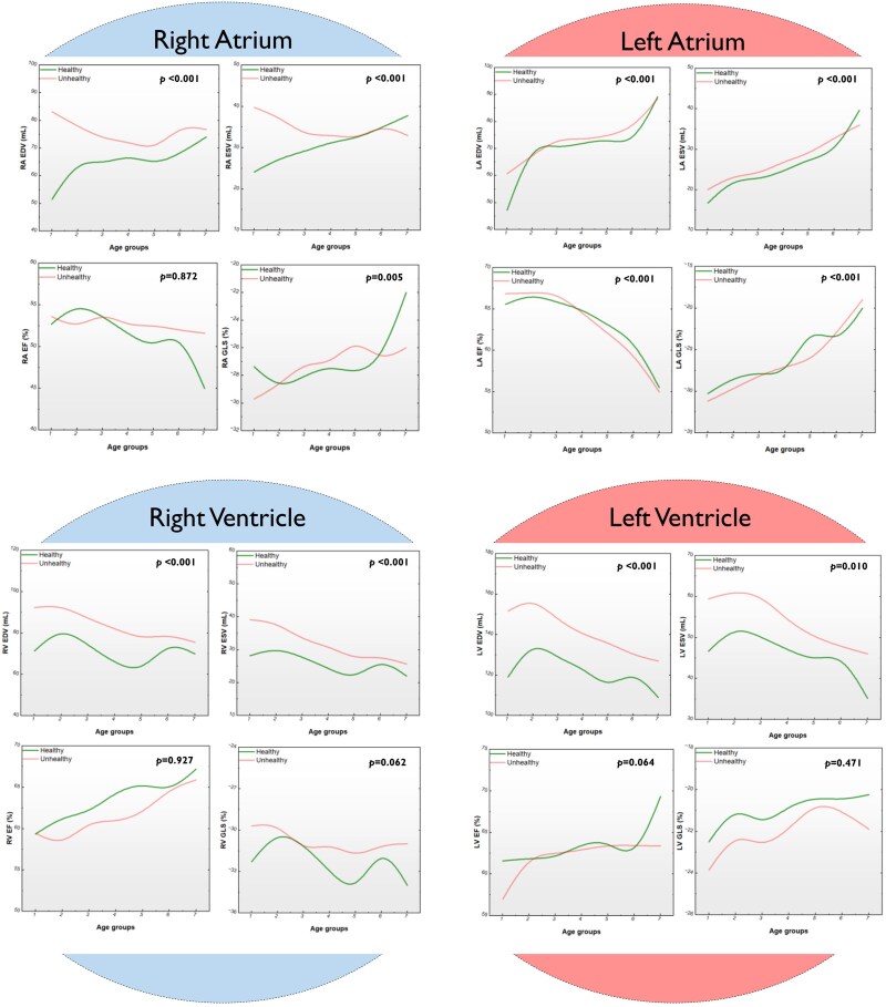
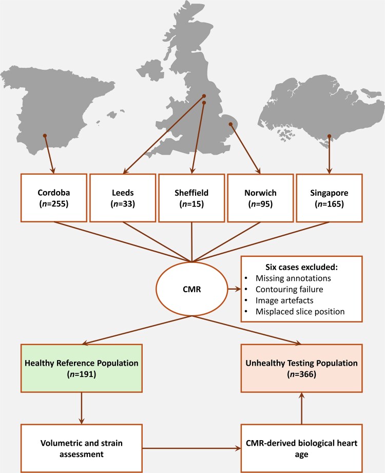
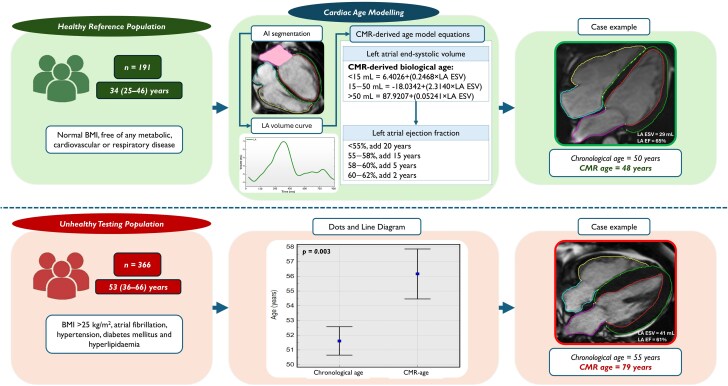
Methods and results: This international, multi-centre, retrospective observational study enrolled 191 healthy individuals with normal body mass index (BMI), free of metabolic, cardiovascular, and respiratory disease as the derivation cohort. Left atrial (LA) end-systolic volume and LA ejection fraction were selected for the final model. The model was validated on 366 patients with BMI >25 kg/m2 and one or more comorbidities [hypertension, diabetes mellitus (DM), atrial fibrillation (AF), and obesity]. In healthy individuals [median age: 34 years, 105 (55%) female], CMR-derived functional heart age was similar to the chronological age [bias: 0.05%, 95% confidence interval (CI): 9.56-9.67%, P = 0.993]. In the validation cohort [median age: 53 years, 157 (43%) female], CMR-derived functional heart age was 4.6 years higher than chronological age (95% CI: 1.6-7.6 years, P = 0.003). Cardiac magnetic resonance-derived functional heart age was significantly higher in patients with hypertension (P < 0.001), DM (P < 0.001), and AF (P < 0.001) than age-matched healthy controls. Moreover, CMR-derived functional heart age was higher than the chronological age in obesity Class I (P = 0.07), obesity Class II (P = 0.11), and obesity Class III (P < 0.001).
Conclusion: This study highlights the time course of structural and physiological changes in the heart during healthy and unhealthy ageing. We propose simple equations that should help communicate subtle changes in heart assessment with ageing.

 求助内容:
求助内容: 应助结果提醒方式:


