Louis Waeckel, Chloé Talon, Mathilde Barrau, Anne-Emmanuelle Berger, Xavier Roblin, Stéphane Paul
{"title":"开发和评估两种全血流式细胞术方案,用于监测接受JAK抑制剂治疗的患者。","authors":"Louis Waeckel, Chloé Talon, Mathilde Barrau, Anne-Emmanuelle Berger, Xavier Roblin, Stéphane Paul","doi":"10.1093/immadv/ltaf006","DOIUrl":null,"url":null,"abstract":"<p><strong>Introduction: </strong>The clinical efficacy of Janus kinase inhibitors (JAKinibs) is highly variable and their safety profiles are poorly understood.</p><p><strong>Methods: </strong>We established two flow cytometry panels for the assessment of two promising leukocyte biomarkers: signal transducer and activator of transcription (STAT) phosphorylation and cytokine receptor expression. We evaluated the first panel, which assesses phosphorylation levels for STAT1, STAT3, and STAT5 after cytokine stimulation, with or without <i>in vitro</i> pretreatment with JAKinibs, in 10 healthy donors. We then evaluated the second panel, which assesses cytokine receptor expression on T cells and B cells, in five healthy donors.</p><p><strong>Results: </strong>Stimulation with interleukin (IL)-2 or IL-7 increased STAT5 phosphorylation in T cells but not in B cells or monocytes. IL-6 stimulation induced STAT3 phosphorylation in monocytes and CD4 T cells and, to a lesser extent, in CD8 T cells, but not in B cells. IL-21 stimulation led to STAT3 phosphorylation in T cells and, to a lesser extent, in B cells, but not in monocytes. Interferon-α stimulation increased STAT1 phosphorylation in all cell types. STAT phosphorylation levels were lower after pretreatment with JAKinibs. A dose-response curve was plotted, confirming the correlation between JAKinib concentration and STAT phosphorylation inhibition. The second panel showed that each cell type displayed a distinct pattern of cytokine receptors expression, and that this pattern might be modified by <i>in vitro</i> treatment with JAKinibs.</p><p><strong>Conclusion: </strong>This preliminary study confirms the utility of flow cytometry for monitoring the biological effects of JAKinibs. Further studies on treated patients are now required to evaluate the clinical value of these two flow cytometry panels.</p>","PeriodicalId":73353,"journal":{"name":"Immunotherapy advances","volume":"5 1","pages":"ltaf006"},"PeriodicalIF":4.9000,"publicationDate":"2025-03-12","publicationTypes":"Journal Article","fieldsOfStudy":null,"isOpenAccess":false,"openAccessPdf":"https://www.ncbi.nlm.nih.gov/pmc/articles/PMC12012447/pdf/","citationCount":"0","resultStr":"{\"title\":\"Development and evaluation of two whole-blood flow cytometry protocols for monitoring patients treated with JAK inhibitors.\",\"authors\":\"Louis Waeckel, Chloé Talon, Mathilde Barrau, Anne-Emmanuelle Berger, Xavier Roblin, Stéphane Paul\",\"doi\":\"10.1093/immadv/ltaf006\",\"DOIUrl\":null,\"url\":null,\"abstract\":\"<p><strong>Introduction: </strong>The clinical efficacy of Janus kinase inhibitors (JAKinibs) is highly variable and their safety profiles are poorly understood.</p><p><strong>Methods: </strong>We established two flow cytometry panels for the assessment of two promising leukocyte biomarkers: signal transducer and activator of transcription (STAT) phosphorylation and cytokine receptor expression. We evaluated the first panel, which assesses phosphorylation levels for STAT1, STAT3, and STAT5 after cytokine stimulation, with or without <i>in vitro</i> pretreatment with JAKinibs, in 10 healthy donors. We then evaluated the second panel, which assesses cytokine receptor expression on T cells and B cells, in five healthy donors.</p><p><strong>Results: </strong>Stimulation with interleukin (IL)-2 or IL-7 increased STAT5 phosphorylation in T cells but not in B cells or monocytes. IL-6 stimulation induced STAT3 phosphorylation in monocytes and CD4 T cells and, to a lesser extent, in CD8 T cells, but not in B cells. IL-21 stimulation led to STAT3 phosphorylation in T cells and, to a lesser extent, in B cells, but not in monocytes. Interferon-α stimulation increased STAT1 phosphorylation in all cell types. STAT phosphorylation levels were lower after pretreatment with JAKinibs. A dose-response curve was plotted, confirming the correlation between JAKinib concentration and STAT phosphorylation inhibition. The second panel showed that each cell type displayed a distinct pattern of cytokine receptors expression, and that this pattern might be modified by <i>in vitro</i> treatment with JAKinibs.</p><p><strong>Conclusion: </strong>This preliminary study confirms the utility of flow cytometry for monitoring the biological effects of JAKinibs. Further studies on treated patients are now required to evaluate the clinical value of these two flow cytometry panels.</p>\",\"PeriodicalId\":73353,\"journal\":{\"name\":\"Immunotherapy advances\",\"volume\":\"5 1\",\"pages\":\"ltaf006\"},\"PeriodicalIF\":4.9000,\"publicationDate\":\"2025-03-12\",\"publicationTypes\":\"Journal Article\",\"fieldsOfStudy\":null,\"isOpenAccess\":false,\"openAccessPdf\":\"https://www.ncbi.nlm.nih.gov/pmc/articles/PMC12012447/pdf/\",\"citationCount\":\"0\",\"resultStr\":null,\"platform\":\"Semanticscholar\",\"paperid\":null,\"PeriodicalName\":\"Immunotherapy advances\",\"FirstCategoryId\":\"1085\",\"ListUrlMain\":\"https://doi.org/10.1093/immadv/ltaf006\",\"RegionNum\":0,\"RegionCategory\":null,\"ArticlePicture\":[],\"TitleCN\":null,\"AbstractTextCN\":null,\"PMCID\":null,\"EPubDate\":\"2025/1/1 0:00:00\",\"PubModel\":\"eCollection\",\"JCR\":\"Q2\",\"JCRName\":\"IMMUNOLOGY\",\"Score\":null,\"Total\":0}","platform":"Semanticscholar","paperid":null,"PeriodicalName":"Immunotherapy advances","FirstCategoryId":"1085","ListUrlMain":"https://doi.org/10.1093/immadv/ltaf006","RegionNum":0,"RegionCategory":null,"ArticlePicture":[],"TitleCN":null,"AbstractTextCN":null,"PMCID":null,"EPubDate":"2025/1/1 0:00:00","PubModel":"eCollection","JCR":"Q2","JCRName":"IMMUNOLOGY","Score":null,"Total":0}
Development and evaluation of two whole-blood flow cytometry protocols for monitoring patients treated with JAK inhibitors.
Introduction: The clinical efficacy of Janus kinase inhibitors (JAKinibs) is highly variable and their safety profiles are poorly understood.
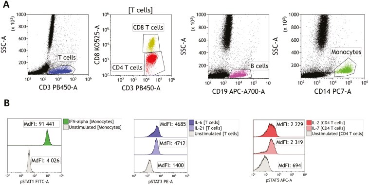
Methods: We established two flow cytometry panels for the assessment of two promising leukocyte biomarkers: signal transducer and activator of transcription (STAT) phosphorylation and cytokine receptor expression. We evaluated the first panel, which assesses phosphorylation levels for STAT1, STAT3, and STAT5 after cytokine stimulation, with or without in vitro pretreatment with JAKinibs, in 10 healthy donors. We then evaluated the second panel, which assesses cytokine receptor expression on T cells and B cells, in five healthy donors.
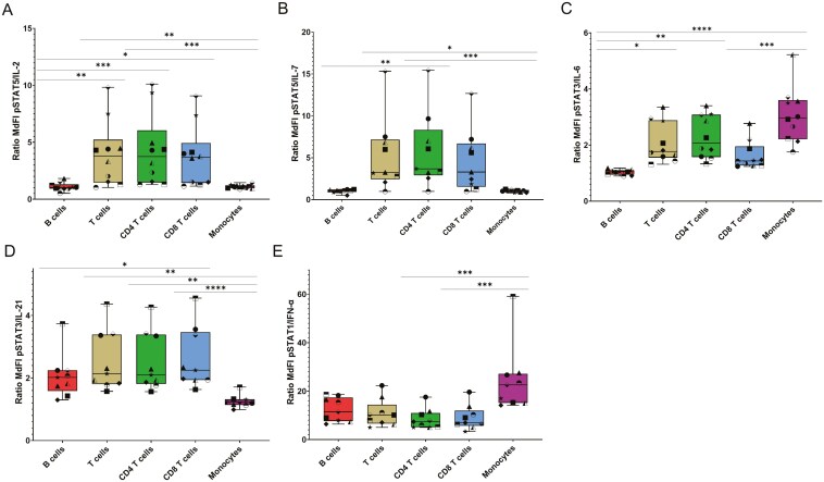
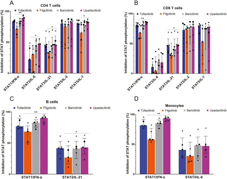
Results: Stimulation with interleukin (IL)-2 or IL-7 increased STAT5 phosphorylation in T cells but not in B cells or monocytes. IL-6 stimulation induced STAT3 phosphorylation in monocytes and CD4 T cells and, to a lesser extent, in CD8 T cells, but not in B cells. IL-21 stimulation led to STAT3 phosphorylation in T cells and, to a lesser extent, in B cells, but not in monocytes. Interferon-α stimulation increased STAT1 phosphorylation in all cell types. STAT phosphorylation levels were lower after pretreatment with JAKinibs. A dose-response curve was plotted, confirming the correlation between JAKinib concentration and STAT phosphorylation inhibition. The second panel showed that each cell type displayed a distinct pattern of cytokine receptors expression, and that this pattern might be modified by in vitro treatment with JAKinibs.
Conclusion: This preliminary study confirms the utility of flow cytometry for monitoring the biological effects of JAKinibs. Further studies on treated patients are now required to evaluate the clinical value of these two flow cytometry panels.

 求助内容:
求助内容: 应助结果提醒方式:


