Saavi Reddy Pellakuru, Ahmed Saad, Karthikeyan P Iyengar, Kapil Shirodkar, Faizul Hassan, David Beale, Rajesh Botchu, Sandeep Velicheti
{"title":"一种识别C2和C3块椎骨的新方法。","authors":"Saavi Reddy Pellakuru, Ahmed Saad, Karthikeyan P Iyengar, Kapil Shirodkar, Faizul Hassan, David Beale, Rajesh Botchu, Sandeep Velicheti","doi":"10.4103/jcvjs.jcvjs_163_24","DOIUrl":null,"url":null,"abstract":"<p><strong>Background: </strong>Congenital block vertebrae (BV) is a common condition resulting from segmentation disorders during embryonic development, leading to the fusion of adjacent vertebrae. BV at C2-C3 (cervical vertebrae 2<sup>nd</sup>-3<sup>rd</sup>) level is the most common segmentation anomaly. Labeling this correctly is the requirement for exact labeling of the spine. Diagnosing BV may not be challenging; however, differentiating BV from the long C2 can be tricky. Our study proposes a novel method of recognizing BV by measuring their height to aid in accurately distinguishing BV from normal vertebrae.</p><p><strong>Methods: </strong>This retrospective study compared C2 vertebral heights between two groups: 50 patients with normal cervical spine magnetic resonance imaging (MRI) and 30 patients with congenital fusion at the C2-C3 levels. Using T2-weighted midsagittal MRI images, the height of the C2 vertebra was measured from the tip of the odontoid process to the posteroinferior part of the vertebra. Data analysis was performed using independent t-tests to evaluate the differences in measurements.</p><p><strong>Results: </strong>The mean C2 vertebral height for the normal cervical spine group was 33.22 mm, while the congenital fusion group exhibited a significantly higher mean height of 45.59 mm. These findings were statistically significant, indicating that a C2 vertebral height exceeding 33 mm is atypical in normal individuals. Our proposed threshold measurement aids in distinguishing between single vertebrae and BV.</p><p><strong>Conclusion: </strong>Our study provides a novel method for assessing C2 vertebral body height to aid in the numbering of cervical spine to enhance diagnostic accuracy in particular in patients with congenital anomalies.</p>","PeriodicalId":51721,"journal":{"name":"Journal of Craniovertebral Junction and Spine","volume":"16 1","pages":"72-76"},"PeriodicalIF":1.3000,"publicationDate":"2025-01-01","publicationTypes":"Journal Article","fieldsOfStudy":null,"isOpenAccess":false,"openAccessPdf":"https://www.ncbi.nlm.nih.gov/pmc/articles/PMC12029387/pdf/","citationCount":"0","resultStr":"{\"title\":\"A novel approach to identifying C2 and C3 block vertebrae.\",\"authors\":\"Saavi Reddy Pellakuru, Ahmed Saad, Karthikeyan P Iyengar, Kapil Shirodkar, Faizul Hassan, David Beale, Rajesh Botchu, Sandeep Velicheti\",\"doi\":\"10.4103/jcvjs.jcvjs_163_24\",\"DOIUrl\":null,\"url\":null,\"abstract\":\"<p><strong>Background: </strong>Congenital block vertebrae (BV) is a common condition resulting from segmentation disorders during embryonic development, leading to the fusion of adjacent vertebrae. BV at C2-C3 (cervical vertebrae 2<sup>nd</sup>-3<sup>rd</sup>) level is the most common segmentation anomaly. Labeling this correctly is the requirement for exact labeling of the spine. Diagnosing BV may not be challenging; however, differentiating BV from the long C2 can be tricky. Our study proposes a novel method of recognizing BV by measuring their height to aid in accurately distinguishing BV from normal vertebrae.</p><p><strong>Methods: </strong>This retrospective study compared C2 vertebral heights between two groups: 50 patients with normal cervical spine magnetic resonance imaging (MRI) and 30 patients with congenital fusion at the C2-C3 levels. Using T2-weighted midsagittal MRI images, the height of the C2 vertebra was measured from the tip of the odontoid process to the posteroinferior part of the vertebra. Data analysis was performed using independent t-tests to evaluate the differences in measurements.</p><p><strong>Results: </strong>The mean C2 vertebral height for the normal cervical spine group was 33.22 mm, while the congenital fusion group exhibited a significantly higher mean height of 45.59 mm. These findings were statistically significant, indicating that a C2 vertebral height exceeding 33 mm is atypical in normal individuals. Our proposed threshold measurement aids in distinguishing between single vertebrae and BV.</p><p><strong>Conclusion: </strong>Our study provides a novel method for assessing C2 vertebral body height to aid in the numbering of cervical spine to enhance diagnostic accuracy in particular in patients with congenital anomalies.</p>\",\"PeriodicalId\":51721,\"journal\":{\"name\":\"Journal of Craniovertebral Junction and Spine\",\"volume\":\"16 1\",\"pages\":\"72-76\"},\"PeriodicalIF\":1.3000,\"publicationDate\":\"2025-01-01\",\"publicationTypes\":\"Journal Article\",\"fieldsOfStudy\":null,\"isOpenAccess\":false,\"openAccessPdf\":\"https://www.ncbi.nlm.nih.gov/pmc/articles/PMC12029387/pdf/\",\"citationCount\":\"0\",\"resultStr\":null,\"platform\":\"Semanticscholar\",\"paperid\":null,\"PeriodicalName\":\"Journal of Craniovertebral Junction and Spine\",\"FirstCategoryId\":\"1085\",\"ListUrlMain\":\"https://doi.org/10.4103/jcvjs.jcvjs_163_24\",\"RegionNum\":0,\"RegionCategory\":null,\"ArticlePicture\":[],\"TitleCN\":null,\"AbstractTextCN\":null,\"PMCID\":null,\"EPubDate\":\"2025/4/1 0:00:00\",\"PubModel\":\"Epub\",\"JCR\":\"Q2\",\"JCRName\":\"OTORHINOLARYNGOLOGY\",\"Score\":null,\"Total\":0}","platform":"Semanticscholar","paperid":null,"PeriodicalName":"Journal of Craniovertebral Junction and Spine","FirstCategoryId":"1085","ListUrlMain":"https://doi.org/10.4103/jcvjs.jcvjs_163_24","RegionNum":0,"RegionCategory":null,"ArticlePicture":[],"TitleCN":null,"AbstractTextCN":null,"PMCID":null,"EPubDate":"2025/4/1 0:00:00","PubModel":"Epub","JCR":"Q2","JCRName":"OTORHINOLARYNGOLOGY","Score":null,"Total":0}
A novel approach to identifying C2 and C3 block vertebrae.
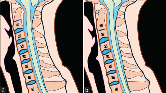
Background: Congenital block vertebrae (BV) is a common condition resulting from segmentation disorders during embryonic development, leading to the fusion of adjacent vertebrae. BV at C2-C3 (cervical vertebrae 2nd-3rd) level is the most common segmentation anomaly. Labeling this correctly is the requirement for exact labeling of the spine. Diagnosing BV may not be challenging; however, differentiating BV from the long C2 can be tricky. Our study proposes a novel method of recognizing BV by measuring their height to aid in accurately distinguishing BV from normal vertebrae.
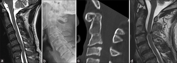
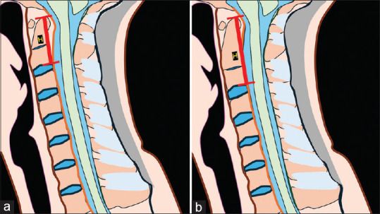
Methods: This retrospective study compared C2 vertebral heights between two groups: 50 patients with normal cervical spine magnetic resonance imaging (MRI) and 30 patients with congenital fusion at the C2-C3 levels. Using T2-weighted midsagittal MRI images, the height of the C2 vertebra was measured from the tip of the odontoid process to the posteroinferior part of the vertebra. Data analysis was performed using independent t-tests to evaluate the differences in measurements.
Results: The mean C2 vertebral height for the normal cervical spine group was 33.22 mm, while the congenital fusion group exhibited a significantly higher mean height of 45.59 mm. These findings were statistically significant, indicating that a C2 vertebral height exceeding 33 mm is atypical in normal individuals. Our proposed threshold measurement aids in distinguishing between single vertebrae and BV.
Conclusion: Our study provides a novel method for assessing C2 vertebral body height to aid in the numbering of cervical spine to enhance diagnostic accuracy in particular in patients with congenital anomalies.

 求助内容:
求助内容: 应助结果提醒方式:


