Romaric Loffroy, Kévin Guillen, Olivier Chevallier, Mohamed Fouad, Emilie Couloumy, Anne Dencausse, Philippe Robert, Sarah Catoen, Anne-Virginie Salsac, Serge Ludwig Aho-Glele, Pierre-Olivier Comby
{"title":"微ct和组织学评价肾动脉栓塞与Glubran®2氰基丙烯酸酯:一项中期随访研究的兔模型。","authors":"Romaric Loffroy, Kévin Guillen, Olivier Chevallier, Mohamed Fouad, Emilie Couloumy, Anne Dencausse, Philippe Robert, Sarah Catoen, Anne-Virginie Salsac, Serge Ludwig Aho-Glele, Pierre-Olivier Comby","doi":"10.1186/s42155-025-00549-8","DOIUrl":null,"url":null,"abstract":"<p><strong>Background: </strong>Cyanoacrylate glues are widely used in interventional radiology as effective embolic agents due to their rapid polymerization and ability to achieve vessel occlusion. Nonetheless, concern remains regarding cast stability and potential recanalization over time. This study used multiple modalities to evaluate the medium-term outcomes of Glubran®2 glue (methacryloxysulfolane and N butyl cyanoacrylate) embolisation in a rabbit renal-artery model.</p><p><strong>Methods: </strong>The left renal arteries of six rabbits were embolized with 12.5% or 25% Glubran®2. In-vivo micro-CT scans were performed immediately after embolisation (M0) and ex-vivo scans and a histological assessment were done at one month (M1). Magnetic resonance imaging (MRI) was done at M1 to assess arterial occlusion and parenchymal changes. Quantitative and semi-quantitative parameters reflecting glue distribution, cast integrity, and tissue response were analysed. Statistical comparisons used non-parametric tests.</p><p><strong>Results: </strong>All six embolisations were completed without complications. Micro-CT at M1 revealed significant cast resorption and fragmentation with both concentrations, but with no evidence of arterial recanalization. MRI and histology confirmed the persistent vascular occlusion with chronic ischemic changes in the renal parenchyma. Compensatory neovascularization from the renal capsule was observed, with no significant differences in histological inflammation between the two concentrations. Glue casts remained within the arterial lumens and were often surrounded by granulomatous inflammation.</p><p><strong>Conclusions: </strong>Glubran®2 was effective for renal artery embolisation, even at a low concentration of 12.5%: despite partial cast resorption, the arteries remained occluded. Micro-CT proved to be a powerful tool for assessing changes in glue casts. Longer-term studies are warranted to further assess vascular remodelling and occlusion durability.</p>","PeriodicalId":52351,"journal":{"name":"CVIR Endovascular","volume":"8 1","pages":"33"},"PeriodicalIF":1.5000,"publicationDate":"2025-04-22","publicationTypes":"Journal Article","fieldsOfStudy":null,"isOpenAccess":false,"openAccessPdf":"https://www.ncbi.nlm.nih.gov/pmc/articles/PMC12014886/pdf/","citationCount":"0","resultStr":"{\"title\":\"Micro-CT and histological assessment of renal arterial embolization with Glubran®2 cyanoacrylate: a medium-term follow-up study in a rabbit model.\",\"authors\":\"Romaric Loffroy, Kévin Guillen, Olivier Chevallier, Mohamed Fouad, Emilie Couloumy, Anne Dencausse, Philippe Robert, Sarah Catoen, Anne-Virginie Salsac, Serge Ludwig Aho-Glele, Pierre-Olivier Comby\",\"doi\":\"10.1186/s42155-025-00549-8\",\"DOIUrl\":null,\"url\":null,\"abstract\":\"<p><strong>Background: </strong>Cyanoacrylate glues are widely used in interventional radiology as effective embolic agents due to their rapid polymerization and ability to achieve vessel occlusion. Nonetheless, concern remains regarding cast stability and potential recanalization over time. This study used multiple modalities to evaluate the medium-term outcomes of Glubran®2 glue (methacryloxysulfolane and N butyl cyanoacrylate) embolisation in a rabbit renal-artery model.</p><p><strong>Methods: </strong>The left renal arteries of six rabbits were embolized with 12.5% or 25% Glubran®2. In-vivo micro-CT scans were performed immediately after embolisation (M0) and ex-vivo scans and a histological assessment were done at one month (M1). Magnetic resonance imaging (MRI) was done at M1 to assess arterial occlusion and parenchymal changes. Quantitative and semi-quantitative parameters reflecting glue distribution, cast integrity, and tissue response were analysed. Statistical comparisons used non-parametric tests.</p><p><strong>Results: </strong>All six embolisations were completed without complications. Micro-CT at M1 revealed significant cast resorption and fragmentation with both concentrations, but with no evidence of arterial recanalization. MRI and histology confirmed the persistent vascular occlusion with chronic ischemic changes in the renal parenchyma. Compensatory neovascularization from the renal capsule was observed, with no significant differences in histological inflammation between the two concentrations. Glue casts remained within the arterial lumens and were often surrounded by granulomatous inflammation.</p><p><strong>Conclusions: </strong>Glubran®2 was effective for renal artery embolisation, even at a low concentration of 12.5%: despite partial cast resorption, the arteries remained occluded. Micro-CT proved to be a powerful tool for assessing changes in glue casts. Longer-term studies are warranted to further assess vascular remodelling and occlusion durability.</p>\",\"PeriodicalId\":52351,\"journal\":{\"name\":\"CVIR Endovascular\",\"volume\":\"8 1\",\"pages\":\"33\"},\"PeriodicalIF\":1.5000,\"publicationDate\":\"2025-04-22\",\"publicationTypes\":\"Journal Article\",\"fieldsOfStudy\":null,\"isOpenAccess\":false,\"openAccessPdf\":\"https://www.ncbi.nlm.nih.gov/pmc/articles/PMC12014886/pdf/\",\"citationCount\":\"0\",\"resultStr\":null,\"platform\":\"Semanticscholar\",\"paperid\":null,\"PeriodicalName\":\"CVIR Endovascular\",\"FirstCategoryId\":\"1085\",\"ListUrlMain\":\"https://doi.org/10.1186/s42155-025-00549-8\",\"RegionNum\":0,\"RegionCategory\":null,\"ArticlePicture\":[],\"TitleCN\":null,\"AbstractTextCN\":null,\"PMCID\":null,\"EPubDate\":\"\",\"PubModel\":\"\",\"JCR\":\"Q3\",\"JCRName\":\"CARDIAC & CARDIOVASCULAR SYSTEMS\",\"Score\":null,\"Total\":0}","platform":"Semanticscholar","paperid":null,"PeriodicalName":"CVIR Endovascular","FirstCategoryId":"1085","ListUrlMain":"https://doi.org/10.1186/s42155-025-00549-8","RegionNum":0,"RegionCategory":null,"ArticlePicture":[],"TitleCN":null,"AbstractTextCN":null,"PMCID":null,"EPubDate":"","PubModel":"","JCR":"Q3","JCRName":"CARDIAC & CARDIOVASCULAR SYSTEMS","Score":null,"Total":0}
Micro-CT and histological assessment of renal arterial embolization with Glubran®2 cyanoacrylate: a medium-term follow-up study in a rabbit model.
Background: Cyanoacrylate glues are widely used in interventional radiology as effective embolic agents due to their rapid polymerization and ability to achieve vessel occlusion. Nonetheless, concern remains regarding cast stability and potential recanalization over time. This study used multiple modalities to evaluate the medium-term outcomes of Glubran®2 glue (methacryloxysulfolane and N butyl cyanoacrylate) embolisation in a rabbit renal-artery model.
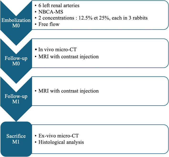
Methods: The left renal arteries of six rabbits were embolized with 12.5% or 25% Glubran®2. In-vivo micro-CT scans were performed immediately after embolisation (M0) and ex-vivo scans and a histological assessment were done at one month (M1). Magnetic resonance imaging (MRI) was done at M1 to assess arterial occlusion and parenchymal changes. Quantitative and semi-quantitative parameters reflecting glue distribution, cast integrity, and tissue response were analysed. Statistical comparisons used non-parametric tests.
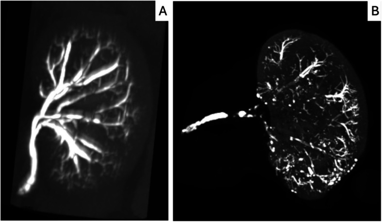
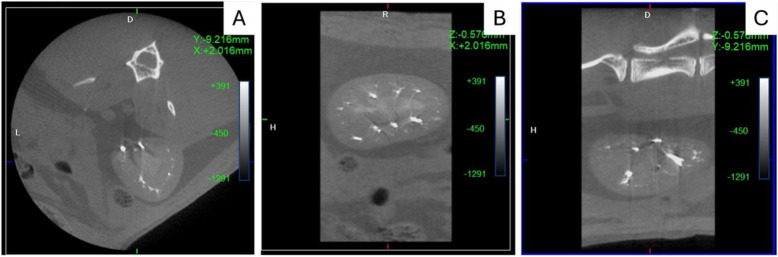
Results: All six embolisations were completed without complications. Micro-CT at M1 revealed significant cast resorption and fragmentation with both concentrations, but with no evidence of arterial recanalization. MRI and histology confirmed the persistent vascular occlusion with chronic ischemic changes in the renal parenchyma. Compensatory neovascularization from the renal capsule was observed, with no significant differences in histological inflammation between the two concentrations. Glue casts remained within the arterial lumens and were often surrounded by granulomatous inflammation.
Conclusions: Glubran®2 was effective for renal artery embolisation, even at a low concentration of 12.5%: despite partial cast resorption, the arteries remained occluded. Micro-CT proved to be a powerful tool for assessing changes in glue casts. Longer-term studies are warranted to further assess vascular remodelling and occlusion durability.
 求助内容:
求助内容: 应助结果提醒方式:


